


IN KERATOCONUS MANAGEMENT, scleral lenses (SLs) are increasingly becoming the “go-to” lens modality for visual rehabilitation. Their ability to correct irregular astigmatism and significantly reduce higher-order aberrations (HOAs) makes them an indispensable tool in optometric practice. However, some keratoconus patients—despite the better visual acuity provided by the improved optics—struggle to adapt to the visual experience provided by scleral lenses, inhibiting their success.
A 35-year-old, long-term spectacle wearer who has moderate keratoconus was fit with SLs. Objective measurements showed significant HOA reduction (Figures 1 and 2): total root-mean-squared (RMS) for the OD improved from 2.61 µm to 0.47 µm, with an improvement in best-corrected visual acuity (BCVA) from 20/25- to 20/20-, while the OS RMS decreased from 3.64 µm to 0.44 µm, with an improvement in BCVA from 20/30-1 to 20/25+. On paper, the optics were excellent; however, she reported “vision is clearer but weird and unnatural,” even after 3 weeks of daily wear and no over-refraction.
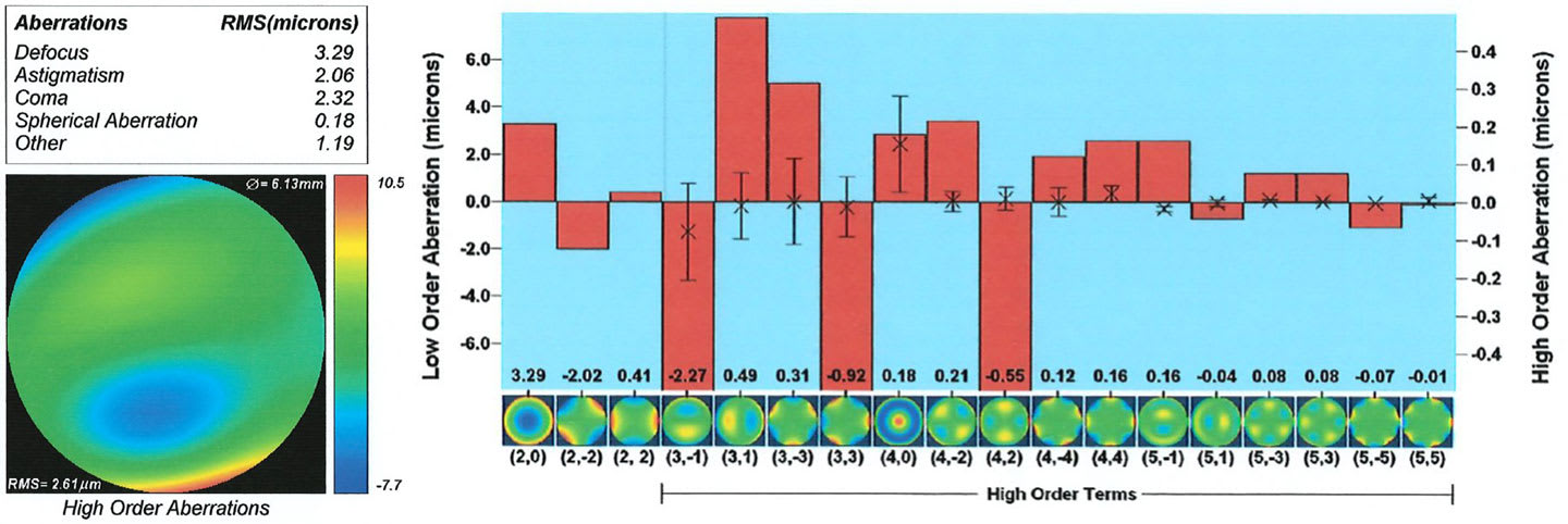
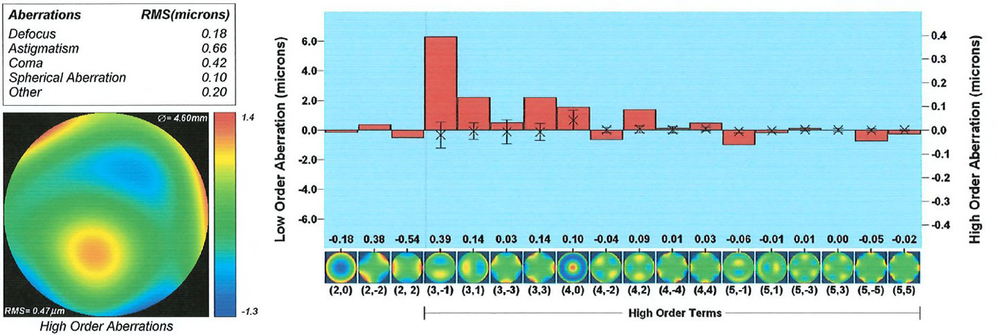
The key insight emerged when comparing her aberrometry with and without the SL. Although the SL reduced the magnitude of her HOAs, it inverted the orientation of her dominant aberration pattern—an expected consequence of neutralizing an irregular cornea with a rigid, rotationally stable surface (Kumar et al, 2021).
For this patient, the brain’s longstanding adaptation to her natural aberration profile made the flipped orientation perceptually disorienting, even if objectively “clear.”
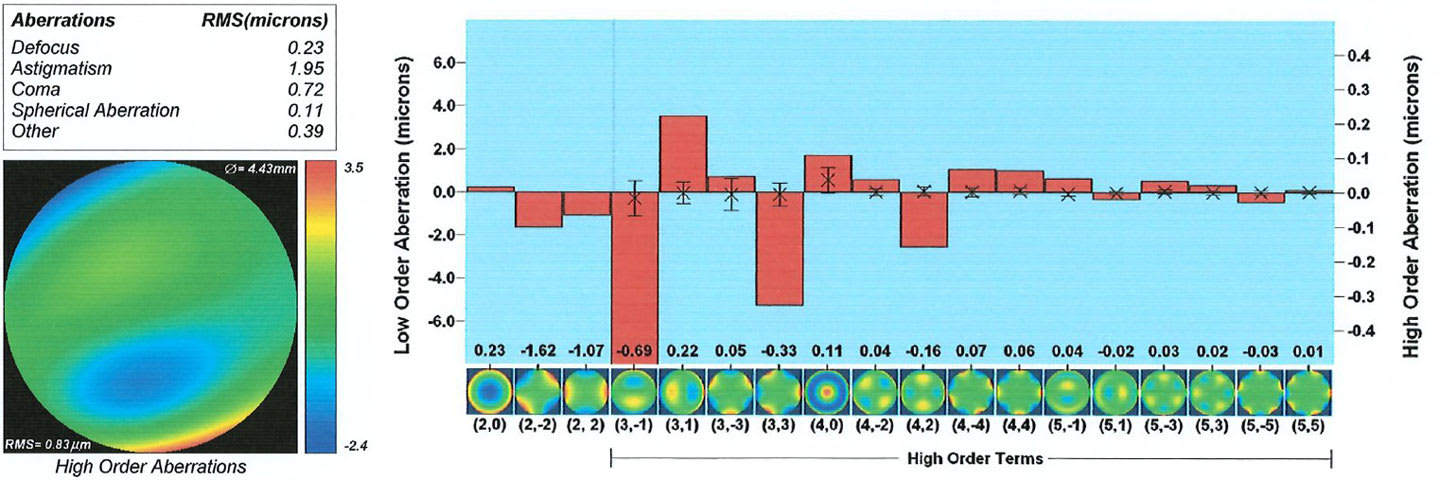
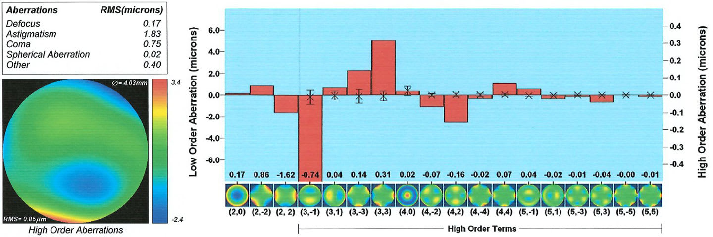
She was subsequently refit into a custom soft keratoconus design with an increased central thickness of 200 µm. These lenses provided partial HOA reduction, but the remaining aberrations preserved the same HOA orientation she experienced with her spectacles (Figures 3 and 4). The result was immediate visual comfort, which increased with time, improved functional acuity over spectacles, and sustained CL success. The outcome was an improved RMS of 0.83 for the right eye and 0.85 for the left eye with a BCVA of 20/20- OD and 20/25+ OS.
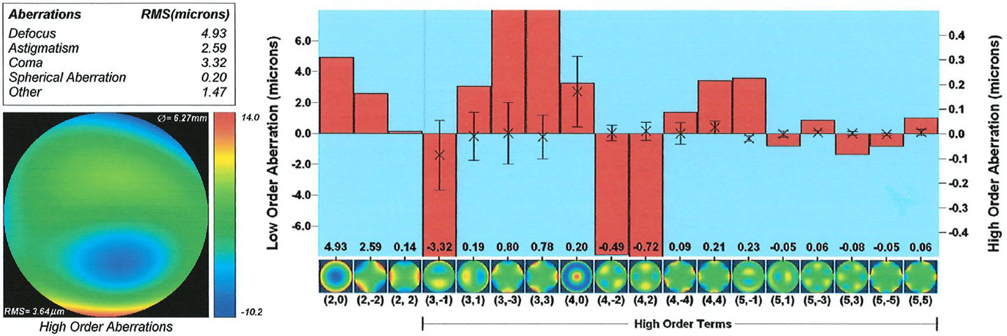
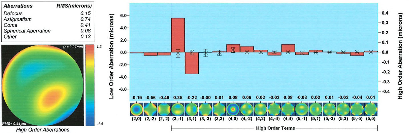
The issue wasn’t inadequate correction or visual improvement but rather disruption to neural compensation of the eye’s aberrations (Artal et al, 2004) (Figures 5 and 6). Unlike SLs, soft contact lenses drape over the corneal surface, leaving the aberration pattern unchanged, and do not conflict with the established neural adaptation present with spectacles.
This case reinforces an important clinical truth: optimal optics do not always equal optimal vision. For some keratoconus patients, especially those deeply adapted to their regular aberration profile, specialty soft lenses may offer the most natural and comfortable visual experience—even when SLs provide superior measured outcomes.
References
1. Kumar M, Shetty R, Lalgudi VG, Khamar P, Vincent SJ, Atchison DA. The effect of scleral lenses on vision, refraction and aberrations in post-LASIK ectasia, keratoconus and pellucid marginal degeneration. Ophthalmic Physiol Opt. 2021; 41:664-672. doi: 10.1111/opo.12802
2. Artal P, Chen L, Fernández EJ, Singer B, Manzanera S, Williams DR. Neural compensation for the eye’s optical aberrations. J Vis. 2004 Apr 16;4(4):281-287. doi: 10.1167/4.4.4