


EVERY ORTHOKERATOLOGY (ortho-k) follow-up tells a story. Between the topography map, visual acuity, comfort, and corneal health, the patient gives you all the clues you need to troubleshoot. But because even a flawlessly designed lens can yield unexpected results, you need a systematic approach when things go sideways.
Connecting the Dots: First, correlate the patients’ subjective symptoms with their axial and tangential difference maps. These maps reveal treatment zone size, centration, and refractive treatment effect. When the map doesn’t match the clinical picture, it’s time to revise the lens.
Small Treatment Zones: Many practitioners intentionally shrink treatment zones when treating rapidly progressing myopia. A meta-analysis confirmed that a smaller back optic zone diameter can enhance myopia control outcomes (Zhou et al, 2024). Sometimes, this drives complaints of glare and halos during night driving or evening sports. Fortunately, increasing the treatment zone size is a straightforward fix that rarely disrupts overall lens fit or centration.
Central Islands: A central island—a steepening in the middle of the treatment zone—usually means excessive sagittal depth. The lens is vaulting too much, which reduces the desired central flattening effect and leaves the patient with fluctuating, poor vision. The fix? Reduce the sagittal depth of the lens. Watch out for “false” central islands. Central superficial punctate keratitis distorts Placido mires, mimicking a central island on topography when the lens fit might actually be fine.
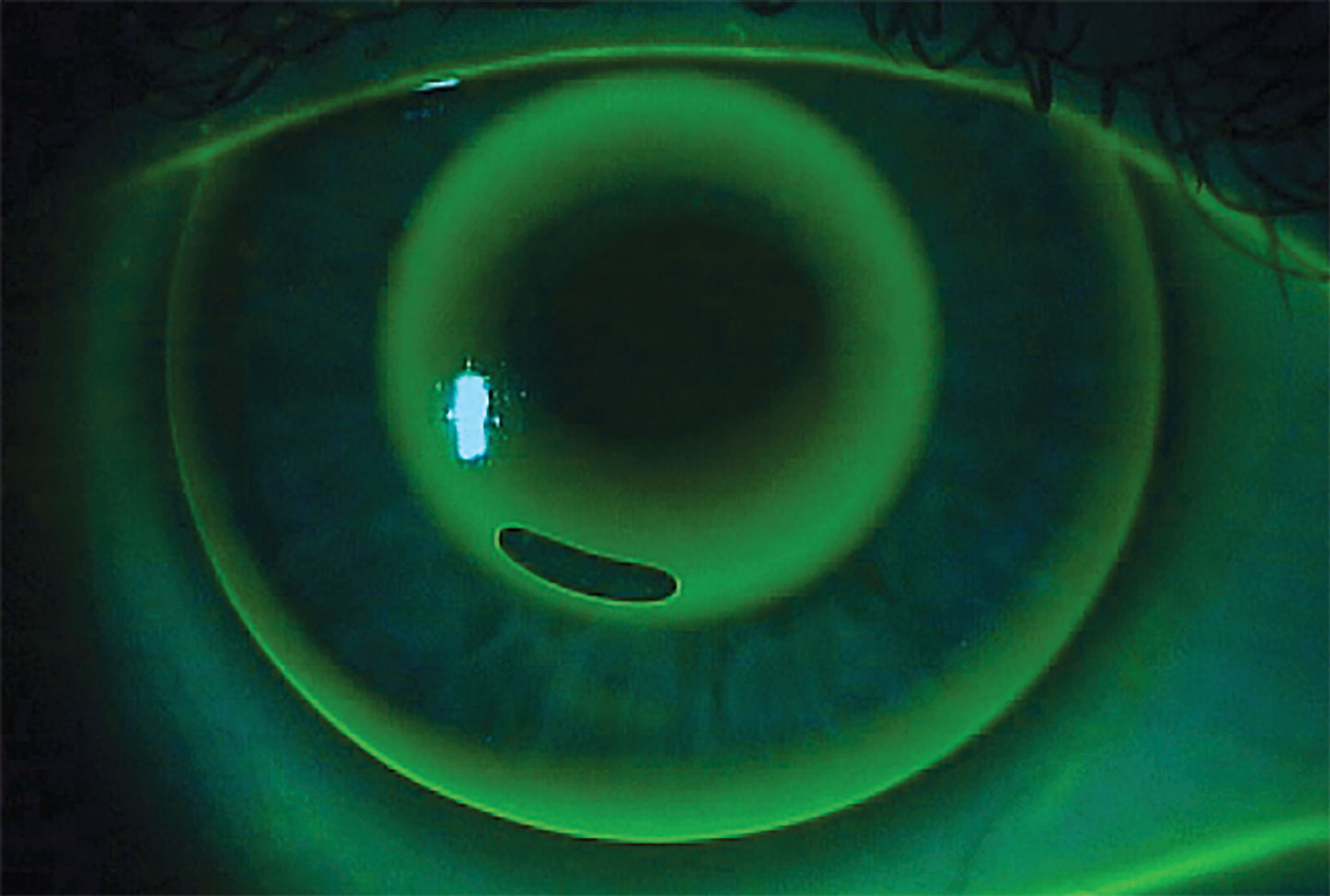
Superior and Inferior Decentration: Inferior decentration gives you the classic “frowny face” map. This usually points to a lens that is too steep or tight. That excessive sagittal depth chokes off tear exchange and causes lens binding and discomfort. Flattening the sagittal depth generally frees up the lens and moves it back into center.
Conversely, superior decentration leads to a “smiley face” map. This means the lens is too flat or loose. Tightening (increasing) the sagittal depth will stabilize the lens, curb excessive movement, and improve comfort.
Lateral Decentration: Nasal or temporal drift usually stems from uncorrected peripheral corneal toricity, asymmetric corneal elevation, or lid forces (like tight eyelids pulling a lens nasally). If you are using a highly customizable design, switching to quadrant-specific modifications is your best bet to lock the lens over the apex.
Undertreatment and Overtreatment: If the lens centers beautifully and moves well, but the refractive target is off, you don’t need to rebuild the lens from scratch. Just adjust the treatment target. Mountford (1997) established that flattening the base curve by roughly 0.10 mm increases the treatment by about 0.50 D without altering the overarching fitting relationship. Don’t rush the revision. Most experienced fitters give the cornea a chance to respond to several weeks of consistent wear before making nonurgent tweaks.
When No Action Is the Best Action: Not every ugly map needs a redesign. If the patient sees well and feels comfortable, and the cornea looks healthy under the slit lamp, step back and observe. Sometimes, the best remedy is leaving it alone.
References
1. Zhou Y, Li H, Hao J, Pan W, Yang Z, Lan W. The efficacy of orthokeratology lenses with smaller back optic zone diameter in myopia control. A meta-analysis. Ophthalmic Physiol Opt. 2024;44(6):1215-1223. doi: 10.1111/opo.13347
2. Mountford J. An analysis of the predictability of orthokeratology. International Contact Lens Clinic. 1997;24(4):129-144. doi:10.1016/S0892-8967(97)00055-2