


IN THE TIMES of ever-advancing new technology for precise fitting of scleral lenses, has the art of diagnostic fitting fallen to the wayside? There are many ways to improve a scleral lens fit by observing fluorescein with the slit lamp: the location, pattern, and rate of movement. There is much more to fitting a lens than assessing central clearance.
The first invaluable diagnostic troubleshooting tip for midday fogging is the application of fluorescein on a settled scleral lens at follow-up visits. By applying fluorescein to the outside of the lens and closely observing the location of excessive tear exchange—or lack thereof—one can determine the area of scleral lens misalignment.
When there is excessive tear exchange during scleral lens wear, patients will complain of foggy or reduced vision and the annoyance of having to clean their lenses frequently throughout the day. Importantly, making the lens tighter or steeper at the periphery is not always the only answer to this problem. Often, looking at the rate and method of fluorescein uptake can tell you whether just the landing zone needs to be adjusted (Figure 1) or if the vaulting chamber itself is misaligned.
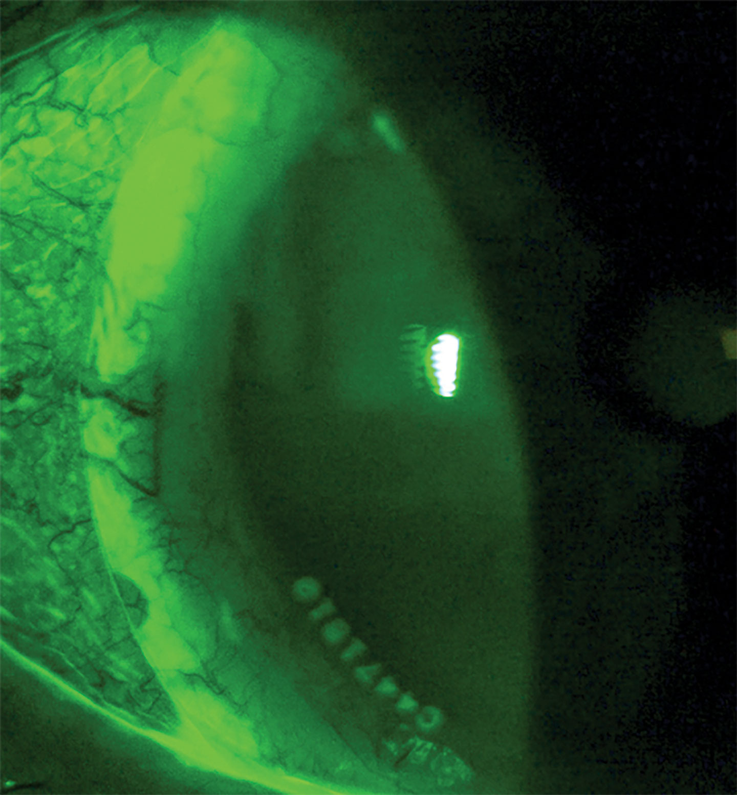
If there appears to be alignment at the edge of a scleral lens or even blanching—despite significant uptake of fluorescein —it is quite likely that the vaulting chamber needs to be extended to align the entire haptic, rather than just steepening the angle of approach as seen in Figure 2.
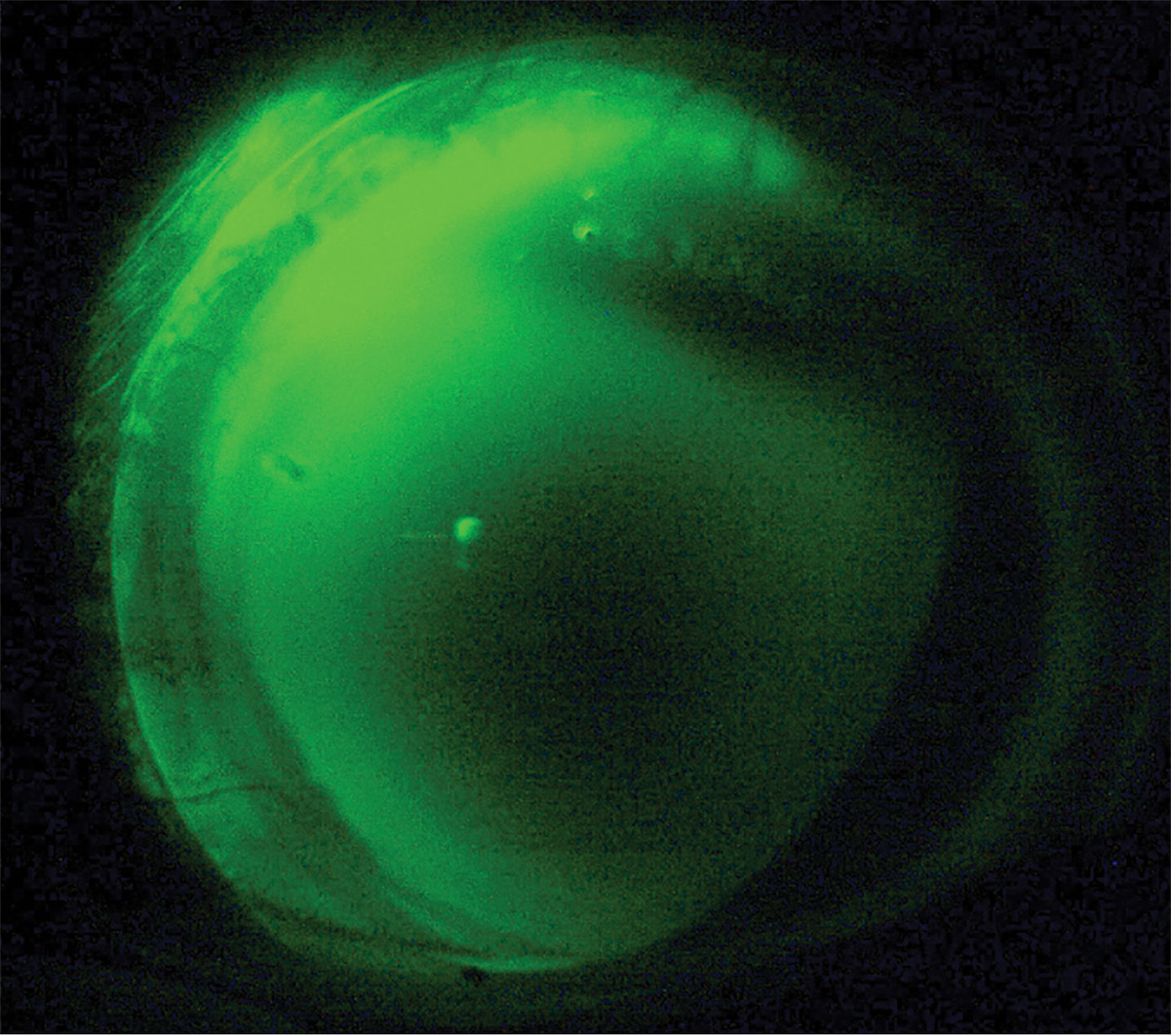
As the patient’s gaze moves around throughout the day, the narrow “toe” or very edge of the lens will lose contact with the sclera briefly, and excessive tear exchange can occur due to the lens overvaulting the limbus. Instead of continuing to steepen the angle of approach further, the more appropriate response is to deepen the vaulting chamber in that quadrant or meridian to better align the entire haptic appropriately, allowing a more stable, comfortable fit.
Another important fitting assessment is to look at the overall picture of the lens fit and limbal alignment with fluorescein when applying a diagnostic lens. Instead of zooming in and assessing just an optic section, the first step should always be to zoom out completely and look at the overall centration and limbal clearance of the vaulting chamber; this is highlighted when fluorescein is instilled into the bowl of the lens while the lens is held in a centered position.
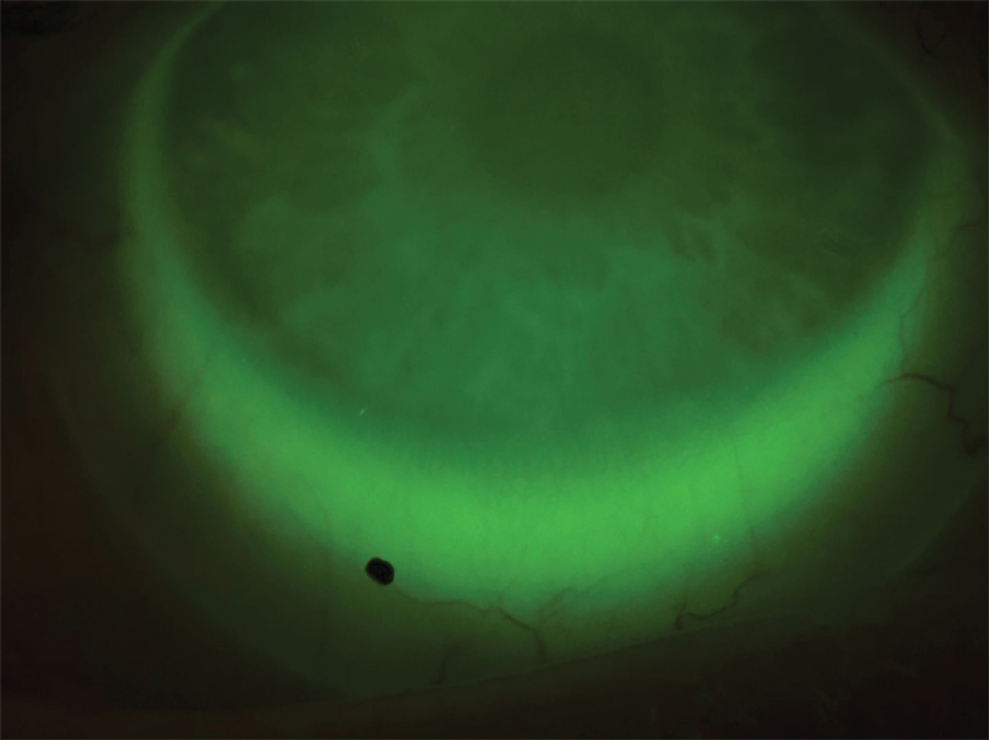
If there is a dark band rather than visible fluorescein around the limbus, that should be the first sign to increase the vaulting chamber or overall diameter of the lens before moving further with the fit. On the opposite end of the spectrum, a lens overvaulting the limbal area in one meridian with decentration (Figure 3) likely needs a dual sagittal depth or bi-elevation lens for proper centration rather than just a standard toric haptic alone.
Even with increasingly sophisticated fitting technology emerging, the use of fluorescein remains a critical and powerful tool when assessing a diagnostic lens—or any scleral lens fit.