contact lens case reports
Help for Screening Abnormal Corneal Topographies
BY PATRICK J. CAROLINE, FAAO, & MARK P. ANDRE, FCLSA
DECEMBER 1998
among the many uses of computer-based corneal mapping systems is screening the abnormal corneal topographies of individuals considering refractive surgery. The major challenge for the equipment is the differentiation between abnormal pathologic corneas, such as keratoconus, and non-pathologic conditions that mimic keratoconus, such as a displaced corneal apex or inferior steepening secondary to contact lens wear.
Pathfinder software, available on Humphrey Instruments' topography systems, evaluates three statistical indices to determine the regularity of the corneal shape: corneal irregularity measurement (CIM), shape factor (SF) and mean toric keratometry (TKM). The three indices are simultaneously displayed on the map with the red portions of the bar indicating the high and low abnormal ranges. The yellow zones indicate borderline ranges and the green indicate normal ranges. The indices for the particular cornea are noted by an arrow above the red bar.
Keratoconus Detection
Patient B.R. is a 32-year-old man interested in corneal refractive surgery. He has a seven-year history of -3.00D OU daily wear planned frequent replacement soft lenses with a visual acuity of 20/20 OU. Routine corneal mapping and Pathfinder analysis correctly identified the patient as a keratoconus suspect, which we later verified at the slit lamp by the presence of faint Vogt's striae OS (Fig. 1).

FIG. 1: Right eye of B.R., identified as a keratoconus suspect |

FIG. 2: S.T.'s left eye with normal with-the-rule astigmatism. |

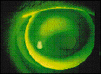
FIG. 3: S.T. fitted with a Rose K design lens, OD 6.75, -4.25D, 8.7mm. |

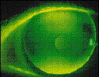
FIG. 4: S.T. fitted with a Boston Envision lens, OS 7.30, -2.00D, 9.3mm.. |
Patient S.T. is a 38-year-old female who was referred to us with the diagnosis of keratoconus OU. Corneal mapping and Pathfinder analysis correctly identified the presence of keratoconus OD, and normal with-the-rule corneal astigmatism OS (Fig. 2). We fitted the patient with RGP lenses (Figs. 3 & 4).
Patrick Caroline is an assistant professor of ophthalmology at the Oregon Health
Sciences University and an assistant professor of optometry at Pacific University.
Mark Andr� is director of contact lens services at the Oregon Health Sciences
University.


