Managing Those Rare Contact Lens Complications
Contact lens wear today is safer than ever, but you still need to know how to recognize and resolve possible problems. This article will help prepare you for the unexpected.
BY WILLIAM D. TOWNSEND, OD
DECEMBER 1998
In the past, all soft lenses had to be enzymed, most contact lens solutions were preserved with thimerosal and heat was the primary means of disinfection. Today's availability of highly gas permeable rigid lenses, as well as disposable and frequent replacement soft lenses, has helped eliminate many of the complications once associated with hard and conventional soft lenses. Many of today's lens materials have much higher Dk values than their predecessors, and there are a wide variety of highly effective, easy-to-use disinfecting systems. Despite these improvements, contact lens related complications continue to present to our offices, often posing a greater challenge for us than fitting the lenses.
History
Regardless of the clinical entity, it's essential that we obtain an adequate history when patients present with a contact lens related complication (Table 1). The quality and thoroughness of the history often determines how well you are able to ascertain the cause of and treatment for the condition. It's particularly important to determine what solution the patient uses with his lenses, if he cleans the lenses regularly and how frequently he replaces them. Be sure you carefully review the time of the onset of symptoms as well as the entire course of the disease. It's very important to determine whether the patient has used any drops or other medications prior to the office visit. In addition to the eye history, the patient's general health history can often reveal potential causes for red eyes and ocular discomfort.
Components of an Adequate History
|
Contact Lens Acute Red Eye (CLARE)
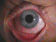
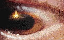
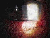
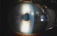
Some patients who overwear their soft lenses or who sleep in lenses not intended for overnight wear may present with frank corneal abrasion or erosion such as appears with hard contact lens overwear syndrome. Other patients may present with symptoms of pain, photophobia, and intense perilimbal injection, but no evidence of frank epithelial abrasion. This condition has been referred to as contact lens acute red eye, or CLARE (Fig. 1). Other associated clinical signs include corneal epithelial edema, conjunctival injection and limbal edema, as well as limbal infiltrates in the peripheral cornea (Fig. 2). The etiology of CLARE is not well understood, but it is believed to be related to the body's immune response to corneal hypoxia. Brien Holden, Ph.D., and others have postulated that the hypoxic corneal epithelium produces a substance that leads to inflammation and leukocyte infiltration. The breakdown of any cell membrane releases membrane phospholipids that are converted into arachidonic acid. This is then converted into prostaglandins by cyclooxygenase and leukotrienes by lipoxygenase. Prostaglandins, particularly PGE2, lead to inflammation, and leukotrienes are chemotactic (attract leukocytes). This may be the final common pathway.
|
|
Other causes of CLARE may include excessively tight-fitting lenses, gram-negative microorganisms, toxicity due to cellular debris trapped beneath a lens and preservative toxicity. It's important to determine the underlying cause of CLARE so that you can prevent its recurrence by making appropriate alterations in lens type, wearing schedule and lens maintenance procedures. Treatment of CLARE is aimed at minimizing patient discomfort. Many of these individuals have anterior chamber activity and benefit from cycloplegia during the initial 24 hours of management. In cases where there is significant anterior chamber activity (cells and flare), topical steroids may be needed for a short time, whereas milder cases may be successfully managed with topical and oral NSAIDs.
It is advisable to use a topical antibiotic in cases where the epithelial integrity is compromised. Joel Silbert, O.D., argues that CLARE can be mimicked by an early bacterial infection and that eyes with CLARE may be more prone to infection. He advocates using a broad-spectrum antibiotic in the initial treatment of this condition. Polytrim (trimethoprim and polymyxin B), an antibiotic with relatively low toxicity, is a good choice for prophylactic management, but should be avoided in patients with known sensitivity to sulfa drugs. The low toxicity of Polysporin ointment makes it an excellent alternative to Polytrim. Regardless of the etiology of CLARE, it's important for patients to discontinue all contact lens wear until the eye is quiet.
Epithelial Splitting
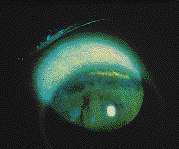
Epithelial splitting (Fig. 3) is a form of noninfectious keratitis that occurs just inside the superior limbus. Like CLARE, its etiology is not well understood, and it is also believed to be due to chronic hypoxia. In a non-contact lens wearer, the superior cornea is hypoxic relative to the cornea within the palpebral aperture because the upper eyelid limits the amount of oxygen available to the cornea beneath it. While this is not a problem for most contact lens wearers, some develop persistent linear staining circumferential to the upper limbus. Differential diagnosis includes a superior limbic keratitis, chlamydial keratitis (adult inclusion keratitis) and corneal abrasion.
|
Epithelial splitting is an indication that the superior cornea needs more oxygen. The lack of conjunctival injection in the quadrant adjacent to epithelial splitting supports the theory that this condition is not inflammatory, but rather due to chronic, low-grade hypoxia. Initial treatment includes discontinuing contact lens wear. Patients with this condition are usually relatively asymptomatic and require minimal pain management. Staining usually resolves within a few days of discontinuing lens wear. Epithelial splitting will usually not recur after refitting the patient in a contact lens with a higher Dk value or higher water content.
Epithelial splitting seems to be more prevalent in toric contact lens wearers and in extended wear patients; it's one more reason why we should routinely remove our patients' contact lenses and evaluate their corneas with sodium fluorescein. The staining in Figure 4 totally resolved after we switched this patient to lenses of an identical design but made of a higher water content material.
|
Giant Papillary Conjunctivitis (GPC)
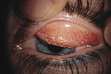
Giant papillary conjunctivitis is a relatively common complication of contact lens wear, although it is also found in individuals with prosthetic eyes, extruded scleral buckles and exposed nylon sutures. Early symptoms and signs include foreign body sensation, itching (especially after lens removal), mild increased mucus production, coated lens surfaces and thickening of the tarsal conjunctiva. As the condition worsens, giant papillae (diameter >1mm) develop and increased lid mass may lead to ptosis of varying degrees. These giant papillae (Fig. 5) often cause contact lenses to ride up and move excessively when the patient blinks. Conjunctival injection and excessive mucus production are also frequently seen in GPC.
|
Mathea Allensmith, M.D., suggested that GPC is caused by a combination of a type 1 and type IV hypersensitivity reaction to the mechanical and immunologic irritation of the tarsal conjunctiva. Degraded proteins, bacteria and mucus form gradually, increasing coatings on the aging surfaces of soft and, to a lesser extent, rigid contact lenses. Allensmith and others have postulated that these coatings are the etiologic factor in GPC development. Histological evaluation of the epithelium in patients with GPC shows mast cells, eosinophils and basophils, all of which the epithelium normally does not contain. In GPC, substantial propria, which normally does not contain eosinophils or basophils, contains large numbers of these cells. Despite the introduction of disposable contact lenses and non-thermal disinfection systems, eyecare practitioners continue to see patients with GPC. This may be due to patients wearing disposable lenses longer than the recommended replacement interval, or due to the tendency of some individuals to coat lenses more rapidly than others.
Giant papillary conjunctivitis must be differentiated from vernal keratoconjunctivitis (VKC), which is unrelated to contact lens wear, but may occur in contact lens wearers. VKC, like GPC, has large papillae with excess mucus production. It occurs most commonly in the spring and fall months and occurs most frequently in adolescent males. Atopic keratoconjunctivitis (AKC) resembles GPC, but it is unrelated to contact lens wear and strongly associated with atopic dermatitis. Table 2 illustrates some signs to help you make the differential diagnosis between GPC, AKC and VKC.
|
Management of GPC begins with replacing coated lenses or, in very severe cases, discontinuing contact lens wear. Stefan Trocme, M.D., showed that treatment with a mast cell stabilizer and frequent replacement lenses allows many patients to continue lens wear. Cromolyn sodium (Crolom) or lodoxamide (Alomide) are key tools for managing GPC and should be prescribed initially on a q.i.d. basis. Patients who complain of itching secondary to GPC often benefit from short-term treatment with a topical antihistamine such as Livostin or Emadine. However, after mast cell stabilizers exert their effect, the antihistamines are rarely needed. Patanol has been shown to have both antihistaminic (H1) and mast cell stabilizing properties. In our experience, Patanol is also an excellent choice as a mono-therapeutic agent for GPC. Topical steroids, while appropriate for initially quieting the lids, have not been shown to be effective in the extended treatment of GPC and should therefore be avoided for long-term management of GPC.
Educate patients with GPC about the cause of their condition, and monitor them frequently for recurrence. Coated contact lenses and excessive lens movement are early signs that should alert you to evert the upper eyelid for signs of GPC.
Solution Hypersensitivity and Toxicity
Despite enormous improvements in contact lens care solutions, solution-related problems continue to plague doctors and patients. These include allergic reactions, toxic keratitis and conjunctivitis, corneal infiltration and nonspecific irritation. Patients with reactions secondary to solution problems can present with a wide variety of symptoms and signs, many of which arise when patients use cleaners, storage solutions and drops that are inappropriate for their lenses.
When contact lens patients call with complaints of redness or irritation, staff members should always instruct them to bring in all of their solutions and drops to the examination. This becomes especially important when the patient has self-medicated with an over-the-counter (OTC) or previously prescribed medication. Many prescription drops and OTC drops are not intended for use with contact lenses and contain preservatives that may become concentrated to a toxic level in soft contact lenses.
Improper use of solutions may also lead to solution-related reactions. When patients fail to completely neutralize hydrogen peroxide-based solutions, or when they confuse their cleaning solution with their lens lubricating drops, they may develop a painful red eye with hazy vision secondary to corneal epithelial loss and edema.
Corneal infiltrates have been identified as a possible result of solution toxicity. Donald Hood, O.D., (Contact Lens Spectrum, Feb. 1994) has suggested that many infiltrates attributed to solution reactions are actually caused by infection or other agents. He found that the true incidence of infiltrates was 0.7% for ReNu and Opti-Free, and 1% for AOSept. When these patients were rechallenged with the same solutions, the recurrence rate was 0% for AOSept and 0.3% for ReNu and Opti-Free. Thimerosal has been known to cause corneal infiltrates but is now rarely used as a preservative in contact lens solutions. Remember, contact lens patients are not immune to other commonly recognized causes of corneal infiltrates, including adenoviral keratoconjunctivitis (Fig. 6), staphyloccal toxin sensitivity and adult inclusion keratoconjunctivitis. These causes should be ruled out prior to treating solution-related reactions.
|
Managing solution-related reactions involves determining and eliminating the causative agent. Sometimes the cause is obvious, as in the chronic use of a product inappropriate for contact lens wear. In cases where the causative agent is less obvious, discontinue all components of the solution regimen and prescribe a new system for the patient. If the patient is very symptomatic, discontinue all lens wear until the eye quiets before instituting a new solution regimen. If infiltrates persist after a patient has started using new solutions, consider the possibility of an infectious process or an allergy to a substance other than the solutions.
Neovascularization
The vast majority of contact lens related corneal neovascularization cases are due to chronic hypoxia and chronic inflammation (Fig. 7). Once you rule out these potential causes of corneal neovascularization, you should investigate other possible etiologies. Corneal neovascularization can result from serious systemic diseases such as syphilis (interstitial keratitis), ocular immune-related conditions such as Terrien's marginal degeneration and Mooren's ulcer, or from a previous episode of infection or trauma. With the introduction of thinner contact lenses with higher Dk values, contact lens associated neovascularization has become less common, but is still seen in clinical practice. Patients wearing contact lenses on an extended wear basis have a significantly higher risk for neovascularization than do daily wear patients. Similarly, patients with chronic inflammatory conditions such as blepharitis or meibomian gland dysfunction have a higher risk of developing corneal neovascularization. When you encounter a patient who is being seen with corneal neovascular changes for the first time, it's important to rule out previous episodes of trauma or infection. This may involve contacting the patient's previous doctors to determine if the neovascularization is new or old.
|
It's important to carefully examine new contact lens patients and first-time contact lens wearers at the time of their initial visit and to record your findings. If you note neovascularization at a later date, the initial findings may be critical in determining the course of management for the patient. If previous records indicate that no vascularization was present when the patient was seen, you must make every effort to determine the cause of the new vessel growth.
Vertical corneal striae or folds (especially later in the day), microcysts and corneal epithelial edema are indications of hypoxia. When a patient with neovascularization presents with these signs, make changes in his lenses or wearing schedule to reduce hypoxia.
Chronic ocular surface disease (OSD) may lead to corneal neovascularization. Signs of OSD include blepharitis, meibomian gland stenosis, inflammation of the meibomian gland orifices and superficial punctate keratitis with peripheral corneal infiltrates (Fig. 8). Patients with OSD present with burning, foreign body sensation and reduced tear film. These symptoms are frequently worse in the morning because inflammatory mediators have been concentrated in the tear film during the night. Increased tear production during the time after the patient awakens dilutes these chemicals and reduces the symptoms. Treatment of OSD includes scrubbing the lids with baby shampoos or commercially prepared pads, instilling a broad-spectrum antibiotic ointment (Polysporin, Bacitracin) and a regimen of heat and meibomian gland expression. Tetracycline and doxycycline alter the esterification process and cause the oils produced by meibomian glands to be thinner and less prone to plugging the gland orifices.
|
Corneal Bacterial and Fungal Keratitis
Bacterial ulceration (Figs. 9a & 9b) is the most severe and sight-threatening clinical entity associated with contact lens wear. All contact lens wearers have a higher risk for ulceration than non-contact lens wearers, but some risk factors are especially significant. Schein et al. (1989) pointed out that extended wear patients have a risk about 10 times greater than users of daily wear lenses. Other factors that increase the risk for bacterial ulceration in contact lens wearers include: diabetes, cigarette smoking, contaminated solutions and cases, use of topical steroids and reduced tear film. Fungal keratitis is very rare and more closely associated with trauma than with contact lens wear, but as Wilson and Ahearn pointed out in the American Journal of Ophthalmology (1996), contact lens wear can be associated with corneal and conjunctival fungal infection.
|
|
When a contact lens patient presents with acute inflammation and evidence of corneal infection, it's important for you to determine whether the patient has corneal ulceration or a less serious condition. Corneal infiltrates may mimic the appearance of an early ulcer. Contrary to what many practitioners believe, staining of an infiltrative lesion is not diagnostic of ulceration. Sterile ulcers may subsequently lose their overlying epithelium and stain with fluorescein. Infectious keratitis usually results in a 360-degree limbal response. In contrast, non-ulcerative infiltrates usually have focal injection in the quadrant where the lesion is located. Patients with bacterial ulcers often present with significant anterior chamber activity or frank hypopion. Non-infectious infiltrates rarely cause significant anterior chamber activity. Infiltrates rarely obscure underlying iris details, but ulceration frequently makes visualization of the iris anatomy difficult.
When examining any patient with a corneal ulcer, a history may provide clues about the nature of the causative organism. It's important to learn what products were used on the lenses and how frequently they were used. Ask patients whether they rinse their lenses in tap water. Be sure to note whether the patient was wearing the lenses on a daily wear or extended wear basis. Equally important is any recent history of eye trauma, exposure to non-chlorinated water or a history of vegetative material in the affected eye. Slit lamp evaluation may be difficult due to photophobia, but it's best to avoid putting any medication, including an anesthetic, in the eye until after the initial evaluation. Be sure you carefully document the location, size and depth of the ulcer and surrounding infiltrate, as well as the presence and severity of any anterior chamber cells or flare. These notes will be important later in judging the effectiveness of the selected treatment regimen. If you elect to manage a suspected bacterial corneal ulcer, proper laboratory procedures must be done prior to initiating treatment. McConnell et al. (American Journal of Ophthalmology, 1992) found that almost half of all southern California ophthalmologists who responded to their survey initiated treatment of bacterial keratitis without first obtaining cultures. The prevalent attitude was that small peripheral ulcers could be treated without a culture, whereas more severe, centrally located ulcers should be cultured and Gram stained before initiating treatment. In a similar study, McLeod et al. (Ophthalmology, 1992) found that, "in general, practice has drifted away from the textbook recommendations of a universal standard for evaluating and treating infectious keratitis." While extensive discussion of this issue is beyond the scope of this work, I advise that you always culture larger, central ulcers, as well as ulcers of any size in monocular individuals and immunocompromised patients. Also culture an ulcer in cases involving trauma, because there is a strong relationship between trauma and fungal ulcers. Any ulcer that doesn't respond to conventional treatment within 48 hours deserves staining and a culture.
Since we're dealing with a very small volume of material, it's better to plate directly onto the solid agar surface. The use of transport media tends to dilute the sample to an unacceptable level. Agar is a polysaccharide extract of red algae to which various types of nutrients are added. There are many different types of media, but for most corneal disease, blood and chocolate agar is all that is required. Blood agar is a general-use medium that contains whole blood cells and will support most bacteria. Some gram negative species, such as Neisseria and Haemophilus, don't grow well on blood agar but will do well on chocolate agar, a nutrient agar in which the red blood cells are lysed. Thayer-Martin agar, a specialized form of chocolate agar, is used when Neisseria infection is suspected. Sabouraud's agar is used to culture fungal organisms. If you have any suspicion of fungal infection, plate on Sabouraud's agar in addition to blood and chocolate agar.
The agent of choice for initial treatment of bacterial ulcers depends on several factors. Monotherapy with a fluoroquinolone may be appropriate for small peripheral ulcers. Ciprofloxacin and ofloxacin have both proven to be effective monotherapy agents, but unfortunately, there are now strains of resistant organisms that don't respond well to these antibiotics. The literature suggests that larger, more severe ulcers should be treated with fortified antibiotics. Consider alternating tobramycin fortified to a concentration of 14 mg/ml with cephalexin fortified to 50 mg/ml every 30 minutes. These agents are toxic when used in these concentrations, so as soon as there's improvement in ulcer size and density, reduce the frequency of dosage. In less severe cases, a topical antibiotic ointment, such as tobramycin, should be instilled in the cul de sac at bedtime. Larger ulcers should be treated with the fortified antibiotic several times throughout the night. Once there's improvement, these treatments can be reduced and eventually eliminated.
Individuals with corneal ulceration should have complete resolution of their condition prior to beginning or continuing contact lens wear. Advise patients to use new contact lenses, cases and solutions since all of these are a potential source of contamination. Patients should understand the importance of contacting you immediately if they experience symptoms indicative of corneal infection.
Eye Health is a Two-Way Street
Contact lens wearers depend on their eyecare providers to keep their eyes healthy, often despite their own unwillingness to comply with recommended lens care recommendations. Many of the problems our patients develop from contact lens wear result from our inability to convince them of the importance of properly cleaning and replacing their lenses. It is incumbent on practitioners and their staff to get this message across to the patient. We must also convince our patients that when they wear contact lenses, it's vital that they notify us if they experience any redness, pain or photophobia.
As eyecare providers, we must be prepared to determine the etiology of contact lens related problems and prescribe appropriate therapy. Ultimately, this is more challenging than deciding which base curve will work best. But it's also much more satisfying and is one more reason why we're considered professionals.
Dr. Townsend is in private practice in Canyon, Texas, and is a consultant at the Amarillo VA Medical Center.


