contact lens case reports
RGPs for Post-LASIK Undercorrection
BY PATRICK CAROLINE, C.O.T., & ROBERT CAMPBELL, M.D.
JANUARY 1998
The theoretical advantages of LASIK include less postoperative pain, less stromal haze, preservation of Bowman's layer and more rapid recovery of vision. Complications are most often flap-related, such a related and include decentration of the flap, microkeratome/laser-induced irregular astigmatism, postoperative flap dehiscence, epithelial ingrowths and flap interface debris. When these complications occur, contact lenses may be required for best corrected visual acuity.
Surgical History
A 39-year-old man with a 20-year history of daily wear PMMA and RGP lenses underwent an uneventful LASIK procedure OD. His preoperative distance Rx was OD -9.00-1.00 x 45, OS -9.00 -1.00 x 151 with visual acuities of 20/20 OU. The target correction (at the plane of the cornea) was -7.00-1.00 x 151, leaving him about -1.00D undercorrected. On the one-day postop visit, we noted fine, putty-like debris in the interface of the flap. The flap was lifted and the debris cleaned and vigorously irrigated with balanced salt solution. On subsequent follow-up visits, only minimal interface debris was noted at six o'clock (Fig. 1).
At six months, the uncorrected visual acuity was stable at 20/40 with a residual refractive error of -1.00-0.50 x 75; 20/20+. The patient was pleased with his uncorrected acuity at near but wanted to periodically wear an RGP lens for full distance correction.
Lens Selection
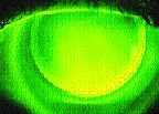
First we obtained a corneal map OD. Central simulated K readings were 37.25@75/ 38.12@164. We chose a diagnostic lens equal to the temporal radius of curvature 4.0mm from center or 41.00D (Fig. 2). Simulated fluorescein revealed 68 microns of central clearance, excellent temporal midperipheral bearing at nine o'clock and slight lens lift off from three to six o'clock over the steeper base of the flap (Fig. 3). The actual fluorescein pattern of the lens closely mimicked the computer simulation (Fig. 4).
Many RGP designs can be used following PTK, PRK and LASIK. With this simple tricurve design, the patient enjoys successful periodic lens wear OD.

FIG. 1: Post-LASIK eye with residual interface debris at six o'clock. |

FIG. 2: Corneal topography OD with slight steepening nasally at the base of the flap. |

FIG. 3: Simulated fluorescein pattern of 41.00D base curve lens. |

FIG. 4: Actual fluorescein pattern of 41.00D base curve lens. |
Patrick Caroline is an assistant professor of optometry at Pacific University and director of contact lens research at Oregon Health Sciences University. Dr. Campbell is medical director of the Park Nicollet Contact Lens Clinic & Research Center, Minnetonka, Minn.


