prescribing for astigmatism
Get the Most Out of Your Corneal Topographer
BY THOMAS G. QUINN, OD, MS
April 1999
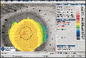
D.K., an active 60-year-old woman, presented wearing 37.5% water content conventional spherical soft lenses (OD +2.50, OS +4.50) for monovision. She complained of having difficulty following her golf ball in flight. Distance vision was 20/25. Spherical refraction over following her golf ball in flight. Distance vision was 20/25. Spherical refraction over the dominant eye (OD), corrected for distance vision, was plano. Refractive findings for the right eye following the removal of her lenses were +2.25 -0.75 x 10², yielding 20/20 acuity. Figure 1 shows axial corneal topography findings. I refit D.K.'s right eye with a 55% water content prism ballasted toric soft lens to enhance distance vision, which provided 20/20 distance acuity and demonstrated good centration and movement.
Although she reported good vision at her follow-up, she noticed reduced comfort after nine hours of wear and irritation and redness after lens removal. The lens again demonstrated good fitting characteristics. After removing the lens, light inferior punctate staining was observed, but no corneal edema was evident. Suspecting hypoxia, I elected to assess the cornea via topography and found inferior corneal steepening induced by toric soft lens wear (Fig.2). I refit her with a lens of higher oxygen transmissibility, which resulted in improved comfort and a return of the topography to baseline.
Topography Map Basics
Corneal topography is an acknowledged tool in managing refractive surgery patients, fitting RGPs and assisting in detecting adverse physiological response in soft lens patients, particularly those wearing torics.
Normal maps show the steepest area located centrally, with a gradual flattening through the mid-region to the peripheral cornea. Steepening in the mid-peripheral region can indicate the presence of corneal swelling.
Nearly all soft torics have some prismatic effect in their design. Whether frank prism or eccentric lenticulation, the inferior portion of the lens is commonly thicker than the superior aspect. This lens thickness, in the materials commonly available today, can cause the cornea to become hypoxic and subsequently cause corneal edema in this inferior region. Therefore, maps showing corneal changes induced by toric soft lens wear will be assymetric, showing greater steepening below the corneal center (Fig. 2).
If lens fitting characteristics are good and there are no overt slit lamp signs,
consider performing topography during follow-up. Although the majority of patients respond
well to today's soft toric designs, topography assures a good physiological response to
contact lens wear.

FIG. 1: D.K.'s right cornea before fitting a toric contact lens.Dr. Quinn
is in group practice in Athens, Ohio, and has served as a faculty member at The Ohio State
University College of Optometry.

FIG. 2: Topography of D.K.'s right cornea after wearing a toric lens for
two weeks.


