contact lens case reports
Lipid Keratopathy: Exploring the Condition
BY PATRICK J. CAROLINE, FAAO, & MARK P. ANDR�, FCLSA
AUGUST 1999
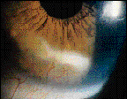
J.K. is a 62-year-old female with a 40-year history of uneventful rigid lens wear. She presented with best corrected visual acuities of OD 20/20 and OS 20/40. External examination of her left eye rerevealed inferior corneal vascularization with a focal band of lipid deposition extending from 5 to 7 o'clock (Fig. 1). Lipid deposition had resulted in significant asymmetry of her corneal topography with the superior cornea at 12 o'clock 47.50D and the inferior cornea significantly flatter at 42.00D (Fig. 2).

FIG. 1: Patient's left cornea with inferior lipid deposition
.

FIG. 2: Corneal mapping OU, note the inferior corneal flattening OS.
The Condition
Lipid keratopathy is the occurrence of fats within the cornea and is usually related to corneal vascularization. Other etiologies include inflammatory anomalies, corneal trauma, dry eye disorders, toxic corneal injuries and nutritional disorders. It may develop suddenly or years after the original diagnosis. The keratopathy appears as dense, whitish-yellow lesions. When the vascularization is confined to a localized area in a compact cornea, the deposition tends to adopt a disc-like shape. When the cornea is swollen, it takes on a fan-like shape, radiating from the ends of the vessels, and in the presence of widespread vascularization, it is diffuse.
The pathogenesis of the lipid deposition may involve increased vascular permeability, excess production of lipids or the tissue's inability to metabolize lipids. Histologically, the material consists of neutral fats, phospholipids and cholesterol crystals.
Primary Management
A serum lipid profile is suggested, especially if the etiology is unclear. If the causative factor is contact lens wear, then all attempts should be directed toward eliminating mechanical trauma to the limbus while increasing oxygen permeability.
Once the keratopathy has been identified and resolved, a portion of the deposition may slowly break up, however, it rarely resolves completely. In this case, the lipid deposition may not have been lens related. The unilateral nature of the condition and the excellent movement and position of J.K.'s present contact lenses might suggest some other past pathology, but her 40-year history of uneventful rigid lens wear can't be overlooked. We eventually refit her with new high-oxygen flux RGPs and instructed her to return for follow-up every 6 months (Fig. 3)

FIG. 3: Patient's OS with an RGP lens.
Patrick Caroline is an associate professor of optometry at Pacific University and an assistant professor of ophthalmology at the Oregon Health Sciences University. Mark Andr� is director of contact lens services at the Oregon Health Sciences University.


