A New Option for Refractive Surgery Patients
By John M. Rinehart, O.D., F.I.O.S.
DECEMBER 1999
Many refractive surgery patients are left with less than ideal results. Find out which contact lens design works well for these patients.
Millions of people undergo refractive surgery each year. Even if the success rate is 90 percent, plenty of refractive surgery patients experience less than ideal results, including undercorrection, overcorrection and irregular corneas. This article explains why rigid gas permeable lenses (RGPs) are the best option to optimize visual acuity for these patients.
Over the years, irregular corneas have challenged every eye care practitioner. Traditional RGP designs typically don't perform well on post-refractive surgery irregular corneas because the central cornea is excessively flat and the peripheral cornea is steep. Many of us have tried reverse geometry lenses on these patients with varying success rates, but the minus-e RGP lens is an additional option for post-refractive surgery patients.
Prior to refractive surgery, the cornea is a prolate asphere, but after surgery, the cornea is an oblate asphere. If an aspheric lens is designed as an oblate, you should be able to create desirable fitting characteristics. To design this lens, you need to know the apical radius of the cornea, the radius of curvature that will produce clearance and the touch areas that are necessary for centration and lens movement. These values are incorporated into the formula in Table 1 to yield the necessary eccentricity value.
| TABLE 1: Eccentricity Value Formula e2 = Rs2 � Ro2 /y2 e = Eccentricity Rs = Sagittal Radius Ro = Apical Radius y = Distance of measured point to apex or 1/2 chord diameter |
Defining Values
The apical radius (Ro) can be either the central keratometer reading or the measurement of apical radius from the corneal topographer. The sagittal radius (Rs) is the measurement in the mid-periphery that will produce the best fitting characteristics for the cornea. As with any lens fit, you want a light touch in the mid-periphery, horizontally and a slight clearance in the vertical meridian. This value can be estimated from computerized topography by measuring the radius of the curvature (3.5mm to 4.5mm) in the temporal quadrant. I use my Reynold's Corneascope to determine the best fitting relationships in the mid-periphery. These values are then applied to the formula in Table 1 to derive the necessary eccentricity. A step-by-step example is described below.
An oblate asphere will result in a negative eccentricity. Provide the lab with the apical radius, the eccentricity and the rest of the lens parameters. Usually, I use a lens diameter of 10.0mm; the optical zone diameter is approximately 0.2mm less than the base curve. Intermediate and peripheral curves will be similar to those of traditional lens designs.
Case History #1
E.M. is a 56-year-old female who underwent refractive surgery six years prior to her visit. Both eyes had four radial incisions, and her right eye had two additional astigmatic incisions. E.M. was unhappy with her unaided visual acuity and wanted to rid herself of eyeglasses. Her unaided acuities were: OD 20/40, OS 20/25 and her subjective refraction was: OD +1.00 - 050 x 025 20/20 and OS +0.50 - 0.75 x 170 20/20. The patient's keratometer readings were: OD 39.50/39.50 @ 090 and OS 40.25/41.00 @ 090.
Computerized corneal topography provides the typical appearance for post-refractive surgery patients (Fig. 1).

FIG. 1: A typical corneal topography view of a post-RK patient.Resulting
Lenses for Case #1
Radius of curvature in the mid-periphery, which appears to provide the best fitting relationships, is (4.1mm from the apex) OD 8.25mm and OS 8.27mm (Table 2). The resulting lenses are illustrated in Table 3.
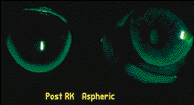
The fluorescein pattern appears similar to an "on K" fit (Fig. 2). The lenses center and move very similarly to a traditional lens design on an eye unaltered by surgery.

FIG. 2: A fluorescein pattern that appears to be an "on K" fit
Because E.W. did not want to wear eyeglasses, the lenses were fit for monovision. Her distance acuity is 20/20 and her near acuity is 20/20. E.M. is able to wear her lenses most of her waking hours. Slit lamp examination showed no disruption of the epithelium and no evidence of corneal edema.
By all standards, this case can be considered successful, resulting in good acuity, comfortable contact lens wear and no disruption of corneal physiology for the patient.
Case History #2
V.G. is a 44-year-old female who had undergone four refractive surgeries in 1996. Each eye has 16 radial incisions and two astigmatic incisions. Neither her distance nor her near acuity was sharp. She also complained that her vision fluctuated, especially when indoors. Her subjective refraction was: OD
-2.25 -0.75 x 035 20/20 and OS - 2.00 - 0.50 x 165 20/20, and her keratometer readings were: OD 39.25/40.75 @ 090 Grade 2 distortion and OS 40.25/40.75 @ 090 Grade 1 distortion.
Photokeratography shows moderate distortion of both corneas (Figs. 3 & 4). The mid-peripheral cornea (4.1mm) is measured as OD 7.98mm (42.25D) and OS 7.84mm (43.00D). The resulting contact lenses can be found in Table 4.

FIG. 3: Photokeratography of V.G. after four refractive surgeries. |

FIG. 4: OU 16 radial incisions and two astigmatic incisions |
A lenticular flange was incorporated into the lens to provide adequate edge thickness. The fluorescein pattern showed apical alignment or possibly a very slight amount of apical clearance (Figs. 5 & 6). There is approximately 1+mm of movement OU. V.G. is presently wearing the contact lenses all of her waking hours without awareness, and both of her corneas are physiologically intact.

FIG 5: V.G.'s fluorescein patterns. |

FIG. 6: OU appears to be alignment or slight apical clearance. |
Case History #3
S.S. is a 61-year-old male who had LASIK performed OU in December 1997. Prior to surgery, his subjective refraction (supplied by the surgeon) was: OD -7.25 - 4.75 x 175 20/30 and OS -7.50 - 3.50 x 002 20/40-50.
In January 1998, the surgeon determined S.S.'s refraction to be: OD -1.25 -4.00 x 016 20/70 and OS +0.25 -4.00 x 120 20/200.
According to the surgeon, S.S. was quite unhappy with these results and was having
difficulty functioning at this level of vision. This is the reason that brought S.S. to my
office in April 1999. He believes that with his contact lenses, both his near and distance
visual acuity are good, but complains that his lenses get cloudy after several hours of
wear. His subjective refraction before the contact lens fitting was: OD
-2.25 -4.00 x 015 20/60 and OS -3.25 -3.00 x 162 20/80, and his keratometer readings were:
OD 39.00/44.00 @ 100 Grade 2 distortion and OS 40.25/43.00 @ 060 Grade 2 distortion.
His current contact lenses decenter nasally and are fixed, and the base curves of both lenses are warped. Based on information supplied to me by S.S., his current contact lenses are as follows:
BC Power Diameter
OD 8.33 -2.00 9.5
OS 7.90 -3.25 9.5
Topography, both photo and video, show both corneas to be very irregular, and it was nearly impossible to get a quality measurement in the mid-periphery to determine the best eccentricity for the new contact lenses (Fig. 7). My best guess for the eccentricity was -0.6 OU. The initial lens parameters are located in Table 5.

FIG. 7: S.S.'s topography before fitting. Note the grossly irregular
corneas.
At dispensing, the contact lenses positioned slightly high and nasally. Acuity through the lenses is OD 20/30 and OS 20/30 -2. After two days of wear, he reported that the comfort with the right lens was great, but that he was experiencing lens awareness with the left lens. The fluorescein pattern showed a near apical alignment, but the lenses continued to decenter nasally OU. In an attempt to improve centration and comfort, I steepened both lenses by 0.50D and increased the diameter of one lens to 10.6mm. This reversed the situation. Now his left lens felt great and he was aware of the right lens. He currently wears the 10.0mm lens on his right eye and the 10.6mm lens on his left eye. His best visual acuities have been 20/30 OD, OS and 20/25+3 OU. S.S. is much happier with the quality of his vision and the comfort of his lenses, which he now wears all of his waking hours.
It would be preferable if the lenses centered better, but considering where we started with the initial fit, the current fit is quite satisfactory. What I like the most is the significant improvement in the corneal topography (Fig. 8). Both corneas appear to be more regular and his central islands have significantly reduced in size.

FIG. 8: S.S.'s topography after fitting. Note the more regular topography.
As you can see from these cases, a minus-e design provides an additional fitting option for post-refractive surgery patients. I have only fit eight refractive surgery patients and one LASIK patient with a minus-e design and all have been successful. However, the diameters I used in these examples may change as more patients are fit.
As of now, the minus-e lens is my lens of choice for post-refractive surgery patients. The fluorescein patterns consistently appear to be "on K" across the entire optical zone, and the lens performs similar to a traditional aspheric lens on a nonoperated eye.
Practice Management Advice
Let me interject some practice management thoughts. These patients paid anywhere from $1,000 to $5,000 to undergo refractive surgery. Therefore, you deserve to be compensated adequately for your efforts in restoring their visual acuity. Because it's extremely likely that the initial lens fit will need to be modified to achieve optimal fitting characteristics, you will need to invest extra time and laboratory costs in these patients. If you are going to supply this higher level of care to your patients, you deserve to have a higher level of compensation.
It's imperative that you inform post-refractive surgery patients that their corneas have been compromised, and that they may not achieve the same results as if their corneas were unaltered. You must be committed to explore every possible design option before discontinuing the care of these patients. If you are not willing to give these patients 100 percent of your time and effort, do the right thing and refer them to someone who is comfortable working with the more sophisticated lens designs.
The next time you choose to fit a post-refractive surgery patient, consider a minus-e design. I think you will be pleased, as will your patient.
Dr. Rinehart currently uses E & E Optics (Van Nuys, California) to manufacture the minus-e design.

TABLE 3Best Fitting Relationships
TABLE 2 OD OS
Base Curve/Eccentricity 8.55/-.5 8.39/-.34
Power +1.00 +3.25 (near)
Diameter 10.0 10.0
Optical Zone Diameter 8.2 8.2
Thickness 0.23 0.28
IC/W 9.00/.3 9.00/.3
Blend 10.00/.2 10.00/.2
PC/W 11.00/.4 11.00/.4
OD OS
Ro = 39.50 (8.55mm) 40.25 (8.39mm)
Rs = 40.87 (8.25 mm) 40.75 (8.27mm)
y = 4.1mm 4.1mm
e2 = 8.252 � 8.552/4.12 8.272 � 8.392/4.12
e2 = 68.06 � 73.10/16.81 68.39 � 70.39/16.81
=-0.2998 = -0.1189
e = -0.547 -0.344
THE EYESSENTIALS
|


