RGP Lens Management of the Irregular Cornea Patient
By Edward S. Bennett, O.D., MSEd, , Michael D. DePaolis, O.D., Vinita
Allee Henry, O.D. and Joseph T. Barr, O.D., M.S.
DECEMBER 1999
Discover the ways in which you can manage patients with irregular corneas easily and successfully.
Managing patients with irregular corneas can be both time-consuming and complex. It is often believed that it either requires too much effort or that these patients are too challenging to manage. The truth is, most of these patients can be managed successfully through the fitting and evaluation of rigid gas permeable (RGP) contact lenses. This article will specifically address the RGP management of three types of irregular cornea patients: (1) keratoconic, (2) penetrating keratoplasty and (3) Laser-assisted in-situ keratomileusis (LASIK).
Management Tips from an Expert
Dr. Edward Bennett uses an approach that incorporates many of the principles used by other eye care practitioners. The common goal is to achieve a three-point touch fluorescein pattern with a spherical RGP diagnostic lens and then to design the lens so that it is consistent with the stage of the keratoconus.
The use of a diagnostic fitting set is imperative when fitting a keratoconic patient with RGP lenses. If you desire an RGP fitting set for your keratoconic patients, it can be obtained from most Contact Lens Manufacturers Association (CLMA) member laboratories. Dr. Bennett's diagnostic set is provided in Table 1.
When fitting a keratoconic patient, the application of a topical anesthetic is beneficial because keratoconic patients tend to be sensitive to initial lens application. Topical anesthetic application will minimize initial lens awareness and reduce chair time. The latter is particularly important since several lenses may need to be attempted prior to obtaining an acceptable fitting relationship. The initial lens should have a base curve radius equal to the steep base curve value, as the Collaborative Longitudinal Evaluation of Keratoconus (CLEK) Study has found this to approximate, on the average, the First Definite Apical Clearance Lens. Do not view the fluorescein pattern immediately after instillation because a false pattern of apical clearance may exist until after several blinks, when bearing is present.
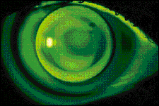
Slit lamp evaluation with both cobalt blue illumination and a Wratten or Tiffen filter will dictate the amount of change in base curve radius that's required. A slight apical clearance fitting relationship will often be present. The base curve can then be changed in 0.50D to 1.00D steps until apical bearing is first observed. At this time, a "three-point touch" or "bull's eye" fitting relationship should be present (Fig. 1).

FIG. 1: A "three-point touch" fitting relationship in a
keratoconic cornea.
Fluorescein evaluation -- Careful evaluation of the peripheral fluorescein pattern is also important to ensure that peripheral seal-off is absent. An alignment fluorescein pattern is not expected with this or with other keratoconic designs; however, good centration is imperative. If the lens decenters inferiorly due to a corneal apex that is greatly displaced in that direction, either a larger diameter can be attempted in an effort to provide better centration or one of the other keratoconic designs discussed in this chapter may be necessary. A Burton Lamp is also valuable for evaluating fluorescein patterns in keratoconic patients, as the greater field of view allows the practitioner to detect if a three-point touch relationship has been achieved more easily.
Designing the lens -- Once three-point touch has been obtained, it's important to design the lens to be consistent with the changes in corneal topography. Generally, the optical zone diameter (OZD) should be decreased in size as the cornea steepens to maintain a well-centered lens. In this philosophy, the OZD is typically equal to the base curve radius (BCR) in millimeters. For example, if the base curve radius is 7.00mm, the OZD will likewise be 7.00mm. Obviously, the OZD can vary depending upon such factors as pupil size, fissure size and lens position.
Multiple peripheral curves (typically 3 to 4) are necessary to correspond with the rapidly flattening mid-peripheral and peripheral cornea. The peripheral curve should generally be flatter and wider than conventional designs to provide greater edge clearance and to prevent peripheral seal-off and lens-to-cornea adherence. This lens design is summarized in Table 2.
| STAGE | AVERAGE "K" VALUE | LENS DESIGN |
| One | <45,00D | Conventional Lens Design |
| Two | 45.00D-50.00D | Overall Diameter = 9.0mm Optical Zone = BCR (mm) Tetracurve Design |
| Three | 50.00D-55.00D | Overall Diameter = 8.8mm-8.8mm Optical Zone = BCR (mm) Tetracurve or Pentacurve design Peripheral Curve Radius = 12.00mm Peripheral Curve Width = 0.3mm-0.4mm |
| Four | >55.00D | Overall Diameter = 8.0mm Optical Zone = BCR (mm) Pentacurve Design Peripheral Curve Radius = 12.00mm Peripheral Curve Width = 3.0mm-0.4mm |
Because almost all lenses ordered will be in minus, if not high minus power, the center thickness should be 0.02mm - 0.03mm thicker than conventional designs to minimize flexure. Contact lenses with powers greater than -5.00D should also be ordered with a plus lenticular or similar peripheral design to minimize edge thickness.
Material selection -- While lens design is the key factor in keratoconus, material selection is also important. Although PMMA and very low Dk (i.e. <20) materials are still in use today because of their ability to correct corneal astigmatism while often providing excellent wettability, it is important to select a material that will not further compromise the cornea via hypoxia.
With the fact that keratoconic lens designs are almost always of minus power and, therefore, thin in design, a fluoro-silicone/acrylate lens material with a minimum Dk value of 30 should provide sufficient oxygen transmissibility, while also providing sufficient rigidity to minimize flexure effects. Extended wear is contraindicated in keratoconus because of the possibility of further compromising an already compromised cornea.
As a result of the irregularity and fragility of the cornea, it is important that keratoconic patients are evaluated on a regular basis. Fortunately, these individuals are often receptive to follow-up care as a preventative measure to ensure the condition is successfully managed.
When the condition is progressing, patients should be evaluated every six months at the minimum. Once it has stabilized, annual examinations, at minimum, should be performed.
The goal in fitting RGPs is not necessarily to obtain a certain fluorescein pattern, but rather to allow adequate wearing time with acceptable comfort, while providing the best vision possible and minimizing tissue insult. Complications are inevitable and a simple guide to problem-solving is provided in Table 3. Problems may result from the lens-to-cornea fitting relationship. If peripheral seal-off is present, the peripheral curve needs to be flatter or wider to increase edge clearance.
| PROBLEM | MANAGEMENT |
| 1. Corneal abrasion/severe swirl staining | C/C CL wear and allow abrasion to heal, then clean posterior surface or consider -0.50D disposable as temporary piggyback. If BCR is too flat, refit with steeper BCR. |
| 2. Paracentral erosion | Most likely due to steep lens with sharp junction. Blend peripheral curve junctions. |
| 3. Peripheral seal-off | Flatten/widen the peripheral curve. |
| 4. Adherence | Flatten/widen peripheral curve or flatten BCR or reduce OZD. |
| 5. Excessive inferior edge lift | Steepen BCR. |
| 6. Poor visual acuity | Select a flatter BCR. |
| 7. Flare | Change to a larger OZD lens. |
| 8. Poor centration | Larger OAD, steeper BCR or use piggyback design (Flexlens, Paragon Vision Sciences, Mesa, Ariz.) |
| 9. Poor comfort | Ensure edges are thin: +lenticular use if indicated. Consider piggyback design; R/0 corneal abrasion. |
If adherence is present, either flattening or widening the peripheral curve, selecting a flatter base curve radius or decreasing the optical zone will typically result in lens movement with the blink. When excessive inferior edge lift is present, the selection of a steeper base curve radius contact lens will reduce this problem.
In certain cases, a special keratoconic lens design specific to the patient's condition (e.g. decentered corneal apex, advanced stage of condition, etc.) may be indicated. There are several lens designs available that could be beneficial in these cases. For summaries of these designs refer to Dr. Shelley Cutler's article, "Managing Keratoconus with Proprietary Designs," (October 1999, CLS).
Penetrating Keratoplasty Treatment
RGP lenses are the most frequently prescribed contact lenses after a penetrating keratoplasty (PK). This is primarily the result of their ability to mask large amounts of irregular astigmatism and provide for an excellent physiologic response. The excellent oxygen transmission, good surface characteristics and flexural resistance of most fluoro-silicone/acrylate lens materials make this category a reasonable choice for post-PK fitting.
Today's post-PK RGP lenses are often 9.5mm-12.0mm in diameter. This larger-diameter philosophy provides for better centration and stability, even in those patients manifesting unusual topographies or graft tilt. RGP optical zones should be as large as possible to facilitate centration and minimize glare, but not so large that harsh bearing, minimal movement or lens adherence results. In situations in which a very large diameter (>10.5mm) is necessary for lens centration and stability, a disproportionately small (<8.5mm) optical zone may be necessary to avoid a steep sagittal depth with attending tear stagnation.
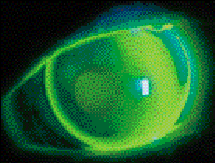
A reasonable starting point for post-PK RGP fitting is to select a base curve that straddles the keratometric measurements. Fluorescein evaluation is then used to refine the base curve to achieve a divided support fit (Fig. 2). In recent years, corneal topography has been recommended as an alternate means for base curve selection. In this approach, the practitioner selects the base curve radius based upon the average dioptric value approximately 3mm from the center of the sagittal topography color map. In patients with atypical topographies, a back surface aspheric lens can be valuable. This often results in good centration and a more uniform, alignment fluorescein pattern.

FIG. 2: A post-PK fluorescein pattern.Keratoconus Problem-Solving
The lens peripheral curve system will depend upon both corneal topography and the fluorescein pattern. Although standard curves are often acceptable, occasionally a very large diameter lens may result in peripheral lens binding, in which case, a series of flatter and wider peripheral curves is warranted. Conversely, in a plateau-type graft, a traditional peripheral curve system might result in excessive edge lift. In these cases, a reverse geometry design is indicated. This lens design is summarized in Table 4.
| Overall Diameter | Large (9.5mm-12mm) |
| Optical Zone | Average (typically below 8.5mm) |
| Base Curve Radius | 1. In between the two K readings 2. If topography is evaluated, select average diptric value 3mm from center of map (sagittal) |
| Peripheral Curve System | Dependent upon topography and fluorescein pattern. If bearing, flatten or widen peripheral curve. If plateau-type graft, use a reverse geometry design. |
LASIK
The contact lens of choice for LASIK patients, if a corrective option is necessary, depends upon the outcome of the surgery. However, if an irregular cornea results, RGP contact lenses are recommended. As with other cases, a fluoro-silicone/acrylate contact lens material is often preferable to a silicone/acrylate.
Selecting an RGP Base Curve
Because the post-LASIK corneal topography is often less prolate in nature, large overall diameters are indicated. This design facilitates lens centration while vaulting the flap, therefore lessening the potential for bearing (Fig. 3). In general, OADs of 9.5mm to 11.5mm are indicated. Given the central flattening associated with myopic LASIK, a large optical zone can cause contact lens adherence or excessive tear pooling, the latter resulting in poor vision, bubble entrapment and hypoxia. To avoid these complications, start with an optical zone that is approximately 2.5mm smaller than the overall diameter, which is considerably smaller than in a standard RGP contact lens design.

FIG. 3: A well-fitting RGP lens after LASIK surgery.
In selecting an RGP base curve for the LASIK patient, practitioners should not rely on post-operative keratometry readings. In most cases, post-operative central keratometric readings will result in a contact lens design that is excessively flat. One method that has been successful is to subtract one-third of the refractive error reduction from the pre-operative flat keratometry for base curve selection. For example, if the patient's pre-operative keratometry readings are 45.00D x 46.00D and the LASIK myopic reduction is 4.50D, then a reasonable initial base curve would be {45.00 - 1/3(4.5)}, or 43.50D.
The transition from central to mid-peripheral cornea in LASIK is often less dramatic than in radial keratotomy (RK) but more so than in surface photorefractive keratectomy (PRK). It is recommended to start with a standard peripheral curve system and modify it accordingly. When evaluating the fluorescein pattern, if excessive bearing is present in the transition zone accompanied by excessive edge lift peripherally, a reverse geometry design is recommended. A summary of RGP lens design parameters for the LASIK patient is provided in Table 5.
| Overall Diameter | 9.5mm-11.5mm |
| Optical Zone | Typically = OAD -2.5mm |
| Base Curve Radius | Flatter than preoperative K reading by 1/4 of total LASIK dioptric reduction |
| Peripheral Curve System | Dependent upon topography and fluorescein pattern. Often can use standard curves. If poor edge clearance, use reverse geometry design. |
RGP Resources
There are a number of useful resources to assist in the fitting of RGP lenses on patients who have irregular corneas. The RGP Lens Institute has several such resources including the RGP Lens Product Guide, which has all of the CLMA member laboratories listed along with address, phone, fax and e-mail information, and all products they manufacture, including keratoconic lens designs. They also have a video entitled "Advanced RGP Fitting" which includes the fitting of bi-toric and irregular cornea patients, and a pocket guide for fitting spherical, bitoric, keratoconic and bifocal lens designs entitled, The RGP Lens Management Guide. These are available by contacting the CLMA at 800-344-9060.
A key factor in the RGP management of the irregular cornea is to manage it yourself. It is often believed that these patients should be referred to the experts (and in certain cases, referral is indicated). However, the key management factor is to use diagnostic lenses and to carefully evaluate the fluorescein pattern. Patience can be a virtue due to the trial and error nature inherent in some of these cases. However, the benefits of fitting these patients with RGPs are many, including helping to improve their self image and enhancing their quality of life. And, of course, the best management option is often the application of RGP lenses.
Dr. Bennett is an associate professor of optometry at the University of Missouri-St. Louis and executive director of the RGP Lens Institute
Dr. DePaolis is in private practice in Rochester, N.Y. and serves as an adjunct faculty member at the University of Rochester School of Medicine and Dentistry and at the Pennsylvania College of Optometry.
Dr. Henry is a clinical associate professor and chief of the Contact Lens Clinic at the University of Missouri - St. Louis School of Optometry.
Dr. Barr is an associate professor at The Ohio State University College of Optometry and a Diplomate in the Section on Cornea and Contact Lenses of the American Academy of Optometry.
THE EYESSENTIALS
|


