Sands of the Sahara: A LASIK Complication
BY DAVID G. KIRSCHEN, OD, PHD, ROBERT LINGUA, MD & CHANG H. KIM, OD
July 1999
Here's an in-depth look at the latest post-operative LASIK complication. Learn how to recognize and manage it in the early stages.
As laser in situ keratomileusis (LASIK) is becoming the predominant refractive surgical procedure of choice for correcting myopia, many surgeons and co-managing practitioners are encountering a new post-operative complication.
Diffuse lamellar keratitis, also known as sands of the sahara (SOS), is a noninfectious condition marked by a powdery infiltrate at the interface between the corneal flap and the stromal bed. The nickname, sands of the sahara, comes from the swirl-like sand pattern that appears in its clinical presentation. Histological studies have identified these infiltrative deposits as polymorphonuclear neutrophils (PMNs), and the pattern of distribution can either be coalesced or diffused at the level of interface.
When the Sands Emerge
Though reports vary, SOS occurs in 1 to 2 percent of patients. It has been reported in unilateral cases occurring in the first or second eye in primary LASIK procedures, bilateral primary LASIK procedures and LASIK enhancement procedures, where the flaps were lifted without the use of a microkeratome. With such a wide variety of occurrences, it is difficult to identify a common denominator that might hint at the etiology of this phenomenon. SOS may present as early as Day 1 or 2 post-operatively, but it is definitely noticeable within the first 7 to 14 days.
Once you diagnose a patient with SOS and you rule out the possibility of an infectious process, begin treatment immediately. Aggressive steroid therapy, close slit lamp evaluation and IOP monitoring are all necessary steps in managing SOS. If the initial treatment is delayed or inadequate, and the inflammation is consequently allowed to progress, then the clinical picture can quickly become more advanced, with the aggregation of inflammatory cells in the interface, which causes a significant drop in the best corrected visual acuity. A hyperopic refractive shift and a change in the topography are both possible. If left untreated, stromal melting due to the production of digestive proteolytic enzyme by neutrophils is also possible. Early recognition and aggressive management of this interface complication should prevent cascading corneal inflammation and other destructive processes, and should ensure an excellent post-operative LASIK outcome.
CASE 1:
Coalesced Infiltrate
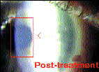
A 47-year-old female had bilateral LASIK to correct her myopia and astigmatism. Four days following the procedure, a slit lamp examination revealed an infiltrate beneath the flap in her left cornea. She was asymptomatic and her visual acuity was normal, at 20/20, as the defect was off the visual axis. Careful slit lamp examination and corneal photography documented a neutrophilic invasion (SOS) at the interface (Fig. 1). Treatment started immediately with 0.1% fluorometholone (Fluor-Op, CIBA Vision) every 2 hours while the patient was awake for a 24-hour period. Then the frequency was decreased to four times per day for 5 days. Follow-up examinations were performed on Days 3 and 6, with the corneal infiltrates completely resolved by Day 6. The steroids were then tapered over a 2-week period. A 3-week follow-up examination yielded a clear cornea and 20/20 acuity (Fig. 2).
|
|
CASE 2:
Diffuse Infiltrate
A 49-year-old male had bilateral LASIK to correct his myopia. His recovery was uneventful until Day 8, when opacities were noted in the interface area on both of his eyes (Fig. 3). His visual acuity on that day was OD 20/40+2, OS 20/50+1. An increase in the corneal haze at the interface was noted on Day 14, but it had remarkably little effect on his vision. The diagnosis of SOS was made and treatment began on Day 15 with Pred Forte, an anti-inflammatory agent by Allergan, four times per day for the first day, every 2 hours for the next day and every three hours for the following day. The anti-infective agent Ciloxan (Alcon) was also prescribed two times per day prophylactically to prevent a secondary bacterial infection during steroid treatment.
On Day 18, there was only a slight improvement, so the Ciloxan was continued twice a day and the Pred Forte was reduced to four times per day. Over the next few weeks, the infiltrate became quite diffuse and on Day 33, the medication was then tapered. Visual acuity at that time was OD 20/25, OS 20/50-2 (20/20 with -1.00 spectacle overcorrection). During the follow-up examination on Day 55, a slight corneal haze was noted that appeared to have little effect on his visual acuity (Fig. 4). He has adapted to the slight monovision correction and is very pleased with the results. He is presently considering the option of an enhancement that will focus both eyes for distance instead of maintaining his current monovision correction.
|
|
Potential Etiologies of SOS
There are several theories pertaining to the etiology of SOS. The four that follow are currently the most popular.
- Sands of the sahara is an exaggerated immune response to lamellar surgery. This theory is thought to be unlikely because undercorrected patients who developed the infiltrate after the initial procedure did not develop SOS after the enhancement, even though a new lamellar cut was made to create a new flap.
- Antigenic sources, including particulate materials from the microkeratome blade and oil residue from the microkeratome motor, result in SOS. This theory is questionable since there are many unilateral presentations of SOS in patients who have undergone a bilateral LASIK procedure using the same microkeratome.
- SOS is a corneal response to thermal injury. Although the laser produces a "cold" beam, it's possible that the energy exchanged at the ablated surface could reach levels high enough to result in thermal damage to the stroma. Some surgeons believe that irrigating the eye with a cold balanced salt solution (BSS) to keep corneal temperature below its normal 94šF range to a cooler 75šF range and pulsing the laser intermittently rather than "blasting away for 50 to 60 shots" keeps the level of thermal injury to a minimum.
- Some believe that ambient temperature and humidity play a role in the etiology of SOS because many cases have occurred in the summer months and in southern states. While climactic conditions may not be solely responsible for SOS, they may certainly be contributing factors.
References are available upon request to the editors at Contact Lens Spectrum. To receive references via fax, call (800) 239-4684 and request document #50. (Be sure to have a fax number ready.)
Dr. Kirschen is in private practice in brea, California. He is full professor of Visual Science and Optometry at the Southern California college of Optometry.
Dr. Lingua is the medical director for TLC Brea Laser Center in Brea, California.
Dr. Kim is the clinical director for TLC Brea Laser Center in Brea, California.


