The Big Picture: Treating the Whole Keratoconus Patient
By Jeffrey J. Eger, O.D., F.I.O.S.
OCTOBER 1999
Looking at the big picture results in improved patient satisfaction and a better contact lens experience.
More than 50 years ago, Dr. Gene Reynolds noticed that some World War II fighters who initially exhibited 20/20 unaided visual acuity became keratoconic after spending time in dark foxholes with bombs screaming overhead. He believed that the extreme emotional stress of fighting and fear of death caused keratoconus. I also subscribe to this theory.
Almost all of my keratoconus patients are very high achievers with a high-stress lifestyle and Type-A personality. They utilize their energy inefficiently. These patients also spend a lot of time on nearpoint tasks, frequently overaccommodating. According to Zimmerman, a direct connection exists between the longitudinal trabecular meshwork fibers and the ciliary body. Each person holds his stress in a different part of the body, and I believe that keratoconics hold their stress in their ciliary muscle. Poor facility occurs due to fatigue and inflexibility in the near focusing system.
In addition, every keratoconus patient I have treated has intraocular pressures between 7mmHg and 12mmHg (measured by Goldmann applanation tonometry). Their eyes appear to be soft because their mires pulse with their heartbeat. I have also discovered that these patients are very high myopes because their eyes are so long. I believe that these factors are related to the problems in keratoconus.
Contact lens practitioners fitting keratoconic corneas need to treat the whole patient, which can be accomplished by discovering the underlying complicating factors of keratoconus, such as emotional stress, divorce, job difficulties or death in the family.
Fitting the Whole Cornea
The rigid gas permeable (RGP) contact lens must be fit meticulously, but on the entire cornea, not just the apex. Make sure you're looking at the healthiest part of the cornea as well as the unhealthy portion. Enhance what's living, ignore what's dying. In a keratoconic patient, the superior part of the cornea is the healthiest, while the inferior cornea is invaginated. You will normally see a dimpled area on the bottom, and the upper apex protrudes out. I fit the superior cornea of a keratoconus patient. The patient is much more comfortable with this type of fit, and contact lenses can be worn upwards of 16 hours a day.
You may often see central staining on many keratoconus patients wearing RGP lenses fit with apical clearance. Many practitioners believe such central staining results from oxygen deprivation. I disagree. Most RGP lens materials today are highly oxygen permeable. Incorrectly fit or apex fit lenses lag down when they fit at the central steep apex. Lenses begin to fit too low and tightly, sealing off the healthiest part of the cornea, the superior cornea. Fresh tears cannot wash behind the lens, and waste products, such as lactic acid, dead cellular debris and carbon dioxide, are trapped. I believe these trapped waste products cause the central staining seen on many keratoconus patients. With an intermediate aligned fit, the lens must attach to the upper lid, and rock on a fulcrum point as the eye blinks. This fit also brings in fresh tears as the patient blinks. Fresh tears re-oxygenate the cornea. When the lens positions up, a bubble of tears squeezes out below the inferior portion of the contact lens, excreting waste products. This helps create a good metabolic pump and homeostasis.
Try fitting flat and intermediate in alignment to the ninth ring of the keratoscope. You'll often see a good, comfortable fit if the lens centers properly, expels tears and is fit in the superior part of the cornea, ignoring the central apex. Patients fit this way say they see better and their lenses feel better. Their corneas look better as well.
Four Patient Management Techniques
1 Flex the accommodative system. I am a case in point stressing the need to work the accommodative system. While in optometry school, I played a lot of sports, but then cut back due to the high reading demand. After this, I noticed I couldn't see the board as well in class.
My acuities were about 20/50 with a refractive error of -1.25D at the time. After an eye examination, I was told I needed eyeglasses that would require a stronger prescription over time. An alternative was vision therapy exercises, such as taking frequent breaks while reading, focusing both near and far, rotation and saccadic exercises and low-plus reading glasses.
I opted not to wear the reading glasses for my refractive error and tried the vision therapy exercises instead. I also knew that after I played sports on the weekends, my vision was fine during the early part of the week. After optometry school, my acuities returned to 20/20 unaided. Today, I am hyperopic and 20/20 unaided at distance. I learned that when the patient is stressed, the periphery closes down. Keratoconus patients are extremely analytical with frequently closed peripheries and overaccommodation.
I recommend to my patients that they take breaks at least every hour or two while working and to look out the window and focus on distance. This exercises the accommodative and motilities muscles. I also prescribe low-plus reading glasses over contact lenses for presbyopic patients.
2 Enhance nutrition. Putting high-quality fuel into our bodies helps them work at peak performance. Low-quality fuel, such as sugar, alcohol, caffeine and fat, results in poor body performance. One patient who returned for follow-up was sure his cornea had changed. Indeed it had. The cornea had flattened, and the lens had tightened. I asked what had happened in his life since our last visit. The patient had stopped drinking coffee following our nutrition discussion. He was previously a 14-cup a day coffee drinker.
He was reluctant to change because his caffeine intake worked for him, but 4 months later, he made the change and felt and slept much better.
3 Begin an exercise program. Exercise not only keeps the body fit, but also reduces stress and rejuvenates the mind, body and soul. Many keratoconus patients are so tense by the end of the day that they're not respirating correctly. I suggest they walk outdoors and look at the sky or the surrounding landscape, not the ground. I see many myopic people walk like they're looking for pennies, which does not promote the good, proper breathing needed to release stress.
If keratoconics don't exercise, the unrelieved stress must go somewhere, and I believe it sits in the ciliary muscle. I also believe the negative energy residing there fatigues the ciliary muscle and causes the cornea to change.
4 Make relaxation a priority. Relaxing also relieves stress and helps the keratoconic patient's accommodative system. Meditation and yoga, with stretching of the neck and shoulders, are excellent relaxation techniques.
Praying is another option. Opening yourself to your faith provides an "out" look. Myopes, including keratoconics, have too deep an "in" look. Looking "out" helps you see more of the "big picture." Relaxing more frequently helps keratoconus patients build up a reserve or buffer against their stress. I find that their corneas don't exhibit as much change and don't require as many frequent refits under high-stress circumstances as they did in the past.
Rather than fitting lens after lens on my keratoconus patients, I challenge them to empower themselves, and to work with me as a team to treat their keratoconus. They need to help change their corneas from soft to stable. If they choose not to do so, their corneas keep changing, their vision is below par, their lenses are uncomfortable and they are not happy with the outcome. Partnering with patients to look at "the big picture" leads to higher success.
CASE 1: Patient K.A. (female, age 21, CPA)
K.A. was diagnosed as needing a penetrating keratoplasty after 2 years of wearing three-point touch and apical clearance-fitted RGPs. Nine months after refitting, her corneas had cleared remarkably, she noticed improved aided and unaided visual acuity and could comfortably wear her intermediate-aligned lenses for more than 15 hours daily. She presently does not require penetrating keratoplasty, and wears +0.75D reading glasses over her contact lenses for near accommodative relaxation and efficiency. The patient is currently wearing Contex OK-2 Airperm contact lenses.
Parameters OS OD
Base curve: 7.6mm 7.7mm
Diameter: 8.3mm 8.7mm
Power: +3.25D +0.25D
CASE 2: Patient A.P. (male, age 30, physics professor)
This spectacle-wearing patient received a keratoconus diagnosis. After he was fit with flatter intermediately-aligned aspheric RGPs and received vision training, he now has 20/30 unaided visual acuity OD, and 20/20 aided and unaided visual acuity OS. The patient wears his contact lenses all waking hours with 20/20 acuity OU.
During the initial fitting, this patient's lenses were regularly refit every 3 to 4 weeks due to extreme corneal changes caused by accommodative stress and a poor exercise and nutrition regimen. He originally opted not to empower himself and partner with the practitioner in his treatment plan. After not accepting an ophthalmology referral for a penetrating keratoplasty, he changed his way of thinking, wore +0.75D reading glasses and initiated an exercise and nutrition plan. The patient is currently wearing Contex OK-4 Airperm Aspheric lenses.
Parameters OS OD
Base curve: 8.05mm 8.5mm
Diameter: 9.5mm 9.3mm
Power: -0.87D +0.50D
CASE 3: Patient D.W. (male, age 40, cardiologist)
This patient was wearing well-fit slightly aspheric rigid gas permeable contact lenses to treat keratoconus when he first presented for care. He was refit with flatter reverse geometry lenses to achieve higher unaided (20/40) and aided (20/20) visual acuities. The patient had good success when partnering with the practitioner in seeing the big picture, walking more upright and breathing normally. However, after 3 months of this success, he decided to stop drinking coffee, later confiding he normally drank 14 cups per day. His corneas consequently flattened, and he required a refit into flatter contact lenses. The patient presently wears +1.00D reading glasses over his contact lenses. He is now currently wearing a Contex Airperm Aspheric 18 lens.
Parameters OS OD
Base curve: 8.28mm 8.45mm
Diameter: 10.5mm 10.3mm
Power: -2.75D -1.25D


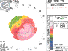
FIG. 1: K.A.'s initial topography. She had been wearing lenses fit by
another practitioner following the three-point touch and apical clearance techniques.

FIG. 2: K.A.'s current topography following 1.5 years of partnering with
her to look at the big picture and refitting

FIG. 3: D.W.'s initial keratoscope findings.

FIG. 4: D.W.'s current keratoscope findings OS after 3 years of looking at
the big picture.

FIG. 5: Keratoconus cornea after treatment showing only trace striae.
THE EYESSENTIALS
|


