


CORNEO-SCLERAL RGPS
Case Report of a Corneo-Scleral RGP Lens
By Todd D. Winkler, O.D., F.I.O.S.
September 1999
This case represents a new treatment option for patients who have undergone corneal transplantation.
Rigid contact lenses have been available in both corneal and scleral designs for approximately the last 50 years. Corneo-scleral rigid gas permeable lenses (RGPs) represent a new category of contact lenses. The Macrolens, which is manufactured by C&H Contact Lens, Inc. (Dallas), is the first such corneo-scleral RGP lens. Incorporating an overall diameter of between approximately 14mm to 15mm, the corneo-scleral lens is larger than a corneal lens, yet smaller than a scleral lens and is supported on the eye by both the cornea and the sclera.
The following case report provides an example where a corneo-scleral RGP contact lens was utilized to provide visual correction to a patient who had undergone a penetrating keratoplasty surgery of his right eye.
Patient History
A local ophthalmologist referred a 37-year-old male to our office for a contact lens fitting. Five years earlier, the patient had suffered an injury to his right eye and consequently required a cornea transplant. The resultant anisometropia rendered eyeglasses an unacceptable form of vision correction, and previous contact lens fitting attempts by other practitioners all failed due to lens intolerance. The patient's chief complaint was intolerable blurred vision in his right eye. After I informed him of a new type of contact lens, the corneo-scleral RGP lens, which offers a reasonable chance of success, he seemed anxious to proceed with the fitting.
Diagnostic Data
The patient's unaided Snellen visual acuity measured less than 20/400 OD and 20/20 OS. His distance refraction yielded the following results: OD -4.75 -2.00 x 175 20/60 and OS +0.25 -0.25 x 90 20/20. Keratometry measurements were as follows: OD 43.75/46.37 @ 90, grade III distortion and OS 44.62/44.25 @ 90.
Biomicroscopy of his right eye revealed a quiet anterior chamber and a clear corneal graft. Biomicroscopy of his left eye and fundus examination of both eyes were unremarkable, as were the results from the remainder of the examination.
Treatment
In complex cases such as this patient's, it's imperative that a trial lens fitting be performed whenever possible. Based upon the keratometry measurements, I selected a trial lens with the following parameters: plano, 7.94mm base curve and an overall diameter of 14.6mm.
I chose a flatter-than-K trial lens because of the considerably large overall diameter. When two lenses have the same base curve but different diameters, the larger lens will possess a greater sagittal area beneath the lens, which produces a steeper fit.
After the lens equilibrated on the eye, I performed a slit lamp evaluation and overrefraction. The trial lens provided good centration and adequate movement, and the fluorescein pattern demonstrated relative alignment centrally with some pooling in the mid-periphery.
Based upon these findings, I had the following Macrolens designed and ordered in the amsilfocon-A material (Dk=45):
| Power | BC | OAD | OZD | CT | PC | |
| OD | 2.50 | 7.94 | 14.6 | 8.0 | 0.17 | aspheric |
At the dispensing visit, the lens displayed the same fitting characteristics as it did during the trial fitting. Acuity in the patient's right eye through the contact lens measured 20/25. Not only was his best corrected acuity improved significantly with the Macrolens, but he was particularly pleased with the increased level of confidence he felt as a result of his improved binocularity and peripheral field of vision.
I dispensed the Macrolens, along with instructions for proper care and lens handling. Subsequent progress examinations were unremarkable.
Post-graft patients need to be monitored closely for suture integrity, neovascularization and graft rejection due to the compromised nature of their corneas. Thus, I instructed the patient to return to our office for further evaluations in 3-month intervals or sooner, if he noted any symptoms of pain, redness or decrease in vision.
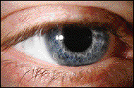
FIG. 1: Corneo-scleral RGP lens on patient's right eye.
|
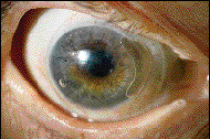
FIG. 2: Lids retracted to show entire lens.
|
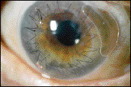
FIG. 3: Magnified view of lens on eye with lids retracted. |
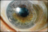
FIG. 4: Entire cornea covered by Macrolens. |
Filling the Void
The corneo-scleral RGP lens has been developed to fill the void left by both soft and rigid contact lenses. Corneo-scleral RGPs provide a level of comfort comparable to soft contact lenses and the better acuity associated with rigid lenses. By utilizing a diameter of between 14mm and 15mm, the same principles of minimal movement and lid interaction associated with soft contact lenses apply. The large overall diameter is advantageous in terms of comfort because the edge of the lens is tucked under both the upper and lower lids (Fig. 1). Thus, the more sensitive lid margins are not in direct contact with the edge of the lens, which eliminates one of the greatest causes of foreign body sensation found with RGPs.
A New Option
Corneo-scleral RGP lenses represent a new treatment option for patients who have undergone corneal transplantation.
The first corneal transplant was performed in 1872. Since then, the number of transplants performed has grown immensely. Over 40,000 corneal transplants are performed in the world each year. The current success rate of the procedure is greater than 95 percent, making it the most successful and the most common of all live-tissue transplants.
The most common indications for this procedure are pseudophakic corneal edema, keratoconus, Fuchs' endothelial dystrophy, stromal dystrophies, post-viral keratitis and corneal scarring secondary to trauma or infection.
High degrees of astigmatism as well as anisometropia, which is often induced by this procedure, can make eyeglasses impractical. However, the complex topography of the post-graft cornea can also make contact lens fitting quite a challenge.
The increased chance of neovascularization with soft contact lenses renders them a less desirable option than RGPs. Because of their greater oxygen permeability as well as their ability to provide better optical correction for irregular corneas, RGPs are the preferred contact lens material to use with corneal grafts.
In many cases, however, corneal RGPs simply cannot be successfully prescribed because of their inability to achieve adequate lens positioning. Rigid lenses frequently decenter to the steepest portion of the cornea. On a typical cornea, this portion is the corneal apex. However, on a post-graft eye, the steepest portion of the graft can be anywhere.
A corneal lens fitted on a post-graft eye is prone to poor centration and has a greater tendency to move off-center, off of the cornea completely or even to fall entirely out of the eye. The larger diameter of a corneo-scleral lens is able to eliminate all of these possibilities more effectively. The larger diameter also provides a broader area of lens alignment and much improved lens stability on an irregular corneal surface (Fig. 2).
When fitting the post-graft eye, the overall diameter should be considerably larger than the diameter of the corneal graft. If the lens is too small, it may bear unnecessarily heavy on the host-graft junction and cause epithelial disruption or may even cause abrasions in the areas of the suture. A typical graft is 7mm to 8mm in diameter. Thus, corneo-scleral lenses are ideally sized for this endeavor (Figs. 3 and 4).
The larger overall diameter of the lens is also more effective in helping to neutralize astigmatic errors. The fenestration serves a dual purpose: not only does it enhance oxygen permeability, but it also helps prevent adhesion of the lens to the cornea.
Just the Facts
If a patient requires rigid lenses, but can't tolerate corneal lenses, and you're not presenting him with the option of corneo-scleral RGP contact lenses, he is possibly being deprived of a visual aid, and in some cases, the only visual aid that could provide him with useful vision.
Corneo-scleral RGP contact lenses represent a new contact lens modality. It's imperative that you become familiar with such designs because the greater the number of tools at your disposal, the greater your likelihood of achieving success for your patients.
Dr. Winkler practices in Cincinnati, Ohio with a special interest in contact lenses. In addition to publishing, he has lectured both nationally and abroad. The author has no commercial interest in either the macrolens or its manufacturer.
Three Types of RGP Lenses:1) Corneal lenses -- all of the pressure is applied to the cornea 2) Scleral lenses -- all of the pressure is applied to the sclera 3) Corneo-scleral lenses -- the pressure is divided between the cornea and the sclera |
THE EYESSENTIALS
|