Identifying Soft Lens Induced Distortion with Corneal Topography
By Kensey S. Inouye, O.D.
SEPTEMBER 1999
Are you aware that corneal topography can be used in the management of soft lens wearers? Find out exactly how.
Used clinically, corneal topographers can provide us with a tremendous amount of information for fitting rigid gas permeable lenses, but they are often overlooked as a tool in managing soft contact lens patients. As part of their software, most topographers have applications that design and simulate a rigid contact lens on the eye being mapped. While articles covering topography and rigid lenses, keratoconus and refractive surgery management appear regularly in the literature, little has been written about its benefit in fitting soft or disposable contact lenses.
What Do You Think?
Marshall Thurber, an authority on W. Edwards Demming's principles on quality and business management, teaches that it's not what you know that can hurt you, but what you know that isn't true. In the past years, contact lens practitioners have been told that a soft contact lens drapes over and conforms to the shape of the cornea and therefore does not distort its shape like a rigid lens can. Many practitioners today still believe this statement, but it may not be true.
To determine whether contact lens practitioners see a need for corneal topography in fitting soft disposable and frequent replacement contact lenses, I asked a few of them at a local gathering to complete a survey according to the following:
Rank your response to the following statements (1= strongly disagree, 2= disagree, 3= neutral, 4= agree or 5= strongly agree):
- Corneal topography is needed in managing refractive surgery patients.
- Corneal topography is needed in managing rigid contact lens patients.
- Corneal topography is needed in managing keratoconus patients.
- Corneal topography is needed in managing soft toric lens patients.
- Corneal topography is needed in managing disposable and frequent replacement lens patients.
Answer yes or no to the following: Do you currently use a corneal topographer in your practice?
Sixty surveys were passed out, and 28 optometrists responded. The results displayed in Fig. 1 represent the mean response of the two groups (users and nonusers) to each statement. Interestingly, there was no significant difference between each group's mean response.
The nonuser group's responses are presented in Fig. 2. This group felt that corneal topography was needed in managing refractive surgery and keratoconus cases, but not in managing soft contact lens cases. Nonusers' opinions were split with regard to topography and rigid lenses.
Using the same graph criterion as above, Fig. 3 shows the response frequencies for the user group. As with the nonuser group, they agreed that topography was needed in managing refractive surgery patients as well as keratoconus patients. Also, like the nonuser group, the user group's decision was split on rigid lenses and they felt that topography was not necessary in managing soft contact lens patients.
Mapping for Hypoxia-Related Changes
In both groups, there was little perceived need for topography in managing soft lens patients. Their responses appear to imply that these optometrists anticipate very little corneal change when fitting soft contact lenses. This may explain why little information has appeared in the literature on topography and soft contact lens management. Although there may be slight corneal change due to the mechanical manipulation of the cornea as with RGPs, corneal changes due to hypoxia should not be ignored.
A Scratch in the Surface
In a recent article addressing oxygen flux through a contact lens, Ken Lebow, O.D., and David Campbell-Burns, O.D., focus on the inconsistencies in measuring and labeling the oxygen transmission (Dk) of a contact lens material and the oxygen transmissibility (Dk/l) of a contact lens design. They describe an example where a lens with an acceptable central Dk/l would not have an acceptable transmissibility because of increased lens thickness in the mid-periphery. This would have resulted in localized corneal edema under the thicker zone.
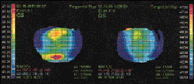
While routinely mapping soft contact lens patients at their initial visit and on subsequent evaluations, I noticed a pattern of mid-peripheral corneal steepening among approximately 90 percent of the patients wearing polymacon lenses. The amount of steepening varied with each individual from 1.00 to 7.00D; however, it was very predictable. If a patient wore a polymacon lens, one could anticipate finding the mid-peripheral steepening. Likewise, if one identified mid-peripheral steepening, the patient usually wore a polymacon lens. The following two cases provide examples of patients exhibiting the corneal distortion with maps taken before and after refitting with mid-water (55%) disposable contact lenses.
Patient #1
Patient #1 is a 23-year-old Caucasian male who originally came in to our office wearing a polymacon disposable contact lens. The left map in Fig. 4 was taken approximately 30 minutes after removing his lens. He did not report any problems with his lenses other than possibly needing a change in his prescription. When questioned further, he reported that he had to rewet his lenses frequently, which he thought was to be expected. The right map was taken 4 days later after refitting him with a 55% water disposable contact lens. The power value in the lower right corner of each map displays the reading at the white cross located at the six o'clock position. Note that the two values differ by 7.32D.
Patient #2
Patient #2 is a 35-year-old Asian male who also presented to our office wearing polymacon disposable contact lenses. His complaints included blurring far vision and lens drying. The left map in Fig. 5 was taken approximately 30 minutes after contact lens removal. The right map was taken after 1 week of wearing a 55% water frequent replacement contact lens. Note the difference in power readings at the cross (4.19D).
In some situations, thin soft lenses made of low water content materials are designed thicker in their mid-periphery to improve handling. In some situations, this added thickness contributes to hypoxia and even corneal distortion. Increasing oxygen flux with the use of a higher water content lens should alleviate the problem. In the cases of the above two patients, this did occur, which leads me to believe that the corneal distortion probably resulted from lens induced hypoxia.
Although this type of corneal change can occur with other materials, polymacon has been known to be the culprit in many of my cases. Because there are numerous factors that affect the oxygen transmissibility of a lens and a cornea's response to reduced oxygen levels (including lens dehydration and lens thickness), you can't assume that a certain lens design and material will or will not cause corneal distortion. You need to evaluate each patient individually and use corneal mapping to monitor for hypoxia-induced corneal changes.
Taking a Closer Look
Should we continue to ignore the benefits of topography in fitting and managing soft contact lens patients? Lenses that we once assumed were acceptable for daily wear may no longer be acceptable. If distortion can occur routinely with disposable lenses, what might we expect with prism ballast toric soft contact lenses? In our survey of practitioners, even those with a topography unit felt that it was not needed in fitting soft lenses. Could this be because they aren't looking for or don't notice corneal changes? Although the corneal distortion described in this article was identified within a small population of soft lens wearers, tools like topographers allow us to identify problems of which we were not previously aware. Keeping our minds open to new ideas and technologies will allow us to find the real truth for the benefit of our patients.
References are available upon request to the editors at Contact Lens Spectrum. To receive references via fax, call (800) 239-4684 and request document #52. (Be sure to have a fax number ready).


FIG. 4: Map showing Patient #1's corneal distortion.

FIG. 5: Map showing Patient #2's corneal distortion.
THE EYESSENTIALS * Sometimes we do not detect problems because we do not look. * Even soft spherical contact lenses can alter corneal topography. |


