Subtle Signs of Sicca -- Advanced Tear Film Assessment
By Jean-Pierre Guillon, BSc, PhD, FCOptom & Graeme Young, BSc,
MPhil, FCOptom, DCLP, FAAO
SEPTEMBER 1999
Here's a review to help you identify contact lens related dry eye problems.
Many of the most challenging complications of contact lens wear have been overcome by more frequent replacement of lenses, less toxic disinfection regimens and higher water content lens materials. Unfortunately, problems relating to dry eye have not been significantly reduced by these changes. In fact, patients' increasing computer usage, coupled with their raised expectations for comfortable contact lens wear, have exacerbated dry eye problems.
An important part of dealing with dry eye complications is diagnosing the exact nature and extent of the problem so that the appropriate course of action can be taken. A recent National Eye Institute Industry workshop (1997) provides a useful framework for categorizing dry eye and lists current methods of diagnosing problems. This workshop, however, was primarily concerned with more severe types of dry eye, whereas the problems most frequently encountered in contact lens practice are only manifest when the tear film is challenged by the presence of a contact lens and are subclinical under normal non-lens-wearing conditions. Hence, many of the standard tests for pathological dry eye (e.g. Schirmer test) are not sensitive enough to identify these marginal cases.
Marginal dry eye sufferers may report a variety of symptoms, ranging from reduced comfort and wearing time to grittiness or other specific dry eye sensations. Their lenses may show desiccation staining following wear with or without an increase in limbal hyperemia.
This article reviews a number of slit lamp signs that are particularly useful in determining possible lens related dry eye problems. These signs are discussed in chronological order, corresponding to the routine of a typical slit lamp examination.
Shallow Tear Meniscus
The height of the inferior tear meniscus is a crude measure of the volume of aqueous in the tear film. Typically, the tear meniscus height varies from 0.1mm to 0.5mm, however, only the shallowest of these can be considered clinically significant. You can view the tear prism with white light, or you can use fluorescein, as long as you take care to instill only a minimal amount. Shallow tear prisms are usually seen in conjunction with a poorly fluorescent tear film (see section on hypofluorescence). Unless you use a measurement graticule, your evaluation of the tear prism depends on your experience and ability to recognize deviations from the norm.
Shallow or Irregular Tear Meniscus
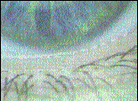
The normal inferior tear meniscus forms a parallel band of tear fluid with a regular upper edge. An irregular tear meniscus forms when there is a relatively low volume of fluid, usually in combination with some other disrupting factor such as an irregular lid margin, irregular bulbar conjunctiva or irregular distribution of lipid secretions along the lid margin. An irregular tear meniscus is therefore highly significant because it often results from two or more of the many risk factors associated with dry eye (Fig. 1).

FIG. 1: Shallow, irregular inferior tear meniscus.
When observing the normal tear meniscus by specular reflection, a bright band of white reflected light may be seen in the middle of the meniscus. This is due to the convex front edge of a relatively high volume tear prism reflecting light back. In the case of a thin tear prism in normal illumination, the white band is extremely narrow because the concave reflecting area is much thinner.
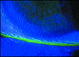
When observing the normal meniscus using the Tearscope Plus (Keeler), a black central line bordered superiorly and inferiorly by bright beams of white light that delineate the meniscus top and bottom can be seen (Fig. 2).

FIG. 2: Black line in tear meniscus.
Conjunctival Folds
Lid-parallel folds in the bulbar conjunctiva adjacent to the temporal conjunctiva become more common with age. Hoh (1995) has shown an association between these lid-parallel conjunctival folds (LIPCOF) and dry eye symptoms. A possible reason for this association is that both dry eye and looseness of the conjunctival tissue are more common with age. However, minor folds in the conjunctiva will be hidden by a deep tear meniscus but will be more evident in the presence of a shallow tear meniscus.
More prominent folds in the conjunctiva will also be hidden by a deep meniscus (Fig. 3) and deprive the tear film of an important reservoir of tear fluid. In severe cases, these folds can contact the cornea, resulting in a foreign body sensation, which can easily be mistaken as a symptom of dry eye.

FIG. 3: Lid-parallel conjunctival folds highlighted by fluorescein.
Meibomian Gland Dysfunction
Without the protective lipid layer that is secreted by the meibomian glands, aqueous within the tear film is free to evaporate. Therefore, a compromised supply of lipid through meibomian gland dysfunction (MGD), threatens the stability of the pre-lens tear film (PLTF). Signs of MGD include froth along the lid margin, hyperemia of the lid margins and waxy excrescences at the meibomian gland openings. Changes to the lid margins, such as hyperkeratinization of the cutaneous margin and rounding of the posterior lid margin due to thickening of the lid, may also be present.
Long-term MGD results in atrophy of individual meibomian glands and irregularity of the lid margins. On digital compression, the meibomian gland secretion may be cloudy or granular (Fig. 4). In more severe cases, it may have a toothpaste consistency (inspissated) which can only be expressed with difficulty. Paugh et al., (1990) showed that a 2-week regimen of warm compresses and lid scrubs can significantly improve tear film stability in problematic contact lens wearers.

FIG. 4: Blocked meibomian glands.
Slow Fluorescein Mixing
The fluorescein break-up test has been shown to have poor correlation with true tear thinning time, however, the instillation and observation of fluorescein mixing at the pre-fitting appointment can be useful indicators. It's best to instill as small a drop of fluorescein as possible in order to prevent flooding the tear film. Instilling fluorescein under the top lid ensures that it follows the normal route of circulating tear aqueous. A relatively thick pre-ocular tear film (POTF) allows for quick circulation of the fluorescein, and the tear film becomes evenly fluorescent within two to three blinks. In the case of a thin tear film, the fluorescein takes more than six blinks to spread across the cornea.
Hypofluorescence
Up to a certain threshold, the thicker a film of fluorescein, the greater the fluorescence will be. Therefore, with a thin POTF, once the fluorescein has been instilled and allowed to spread, the tear film will show relatively poor fluorescence. In extreme cases, the fluorescence of the tear film may only be visible in the tear meniscus.
Clearly, the level of fluorescence depends on the amount of fluorescein instilled into the eye. Another confounding factor is that, in extreme cases, a thick lipid layer can impede the transmission of blue light and give the false impression of hypofluorescence. However, by consistently using a small amount of fluorescein you will eventually learn to distinguish normal levels of fluorescence from abnormal levels.
Fluorescein Banding
Horizontal bands of hypofluorescence, visible as black lines, indicate areas of tear film thinning and possible subsequent epithelial desiccation. These troughs in the tear film form close to the lid margin due to its proximity to the tear meniscus, which, in the case of a small meniscus, induces a relatively high capillary force. The sign is even more noteworthy if the band is wide and irregular and is frequently associated with other diagnostic signs of dry eye.
In non-lens wearers, bands of hypofluorescence may be present, without leading to corneal staining or discomfort. However, because soft contact lenses always support a thinner tear film than the cornea, there is greater potential for staining.
The absence of PLTF in these areas allows localized evaporation, dehydration and ultimately, epithelial desiccation to occur. Horizontal bands can also form in a position that corresponds to the lowest extent of an incomplete blink and, again, result in desiccation.
Desiccation Staining
A proportion of non-lens wearers show some epithelial staining, however, desiccation
staining due to a tear film surfacing anomaly can be distinguished by its location and
pattern. Desiccation staining is most commonly located 1-2mm above the lower lid margin,
although in the presence of an incomplete blink, it may be found in the central cornea
(Fig. 5). In both cases, the staining forms a horizontal band of punctate staining
corresponding to the bands of hypofluorescence discussed earlier.

FIG. 5: Desiccation staining.
Thin Aqueous or Lipid Layer
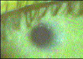
The lipid layer is just visible using a slit lamp and specular reflection. However, a wider, more revealing image of the lipid layer can be seen using the Tearscope. The thickness of the lipid layer can be gauged from the appearance of the interference pattern. The thickest type of lipid pattern resembles oil on water, while the thinnest lipid films are fainter and show a marble-like pattern (Fig. 6). Although the normal POTF always supports a lipid layer, this is not always the case with the PLTF. Patients who show the thinnest type of pre-ocular lipid film often show no lipid film in their PLTF and are likely to experience evaporative dry eye.

FIG. 6: Thin pre-lens tear film lipid layer
In the presence of a thick lipid layer, interference patterns in the pre-lens aqueous layer are not visible, however, with a thin or absent lipid layer, Newton's ring-type interference fringes are visible. In contrast to the lipid interference fringes, Newton's ring-type interference fringes are dark and light and form in lines of equal thickness (Fig. 7). This type of aqueous layer, visible on Tearscope examination, suggests a thin, relatively unstable tear film. Another warning sign of a thin aqueous layer is specks of dust in the tear film that fail to clear with a blink. Where the aqueous layer is thin, these dust particles are less easily washed away into the tear prisms.

FIG. 7: Visible fringes in aqueous layer in the absence of lipid layer.
Rapid Drying Time
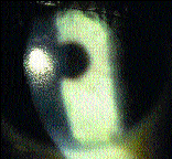
The above signs are indicative of potentially troublesome contact lens wear. With soft lenses, the acid test indicates whether the lens itself can support a stable tear film. A useful diagnostic test, therefore, is to observe the PLTF after an appropriate settling period. An unstable tear film might be evident after only a few minutes, but in some cases, a longer period of time (e.g. >30 minutes) is required. The presence of a lipid layer on the PLTF is an encouraging sign, but the length of the drying time is the most critical feature. Logically, the drying time should be longer than the normal inter-blink period. In practice, the effect and clinical significance of pre-lens drying may depend on a number of factors, but primarily depends on whether front surface drying translates into drying of the post-lens tear film and corneal desiccation.
Spot drying, particularly if it occurs repeatedly in the same position, is more damaging than uniform thinning of the tear film (Fig. 8). A prolonged drying time is less relevant if incomplete blinking fails to rewet inferior areas of dryness. Pre-lens drying will be less problematic with a thick, mobile lens or a lens that is manufactured from a dehydration-resistant material.

FIG. 8: Pre-lens drying with uniform thinning of the tear film.
Conclusion
The conventional methods of testing for dry eye are insufficient in normal contact lens practice. Good observational skills and intelligent interpretation of the subtle signs of marginal dry eye are required to successfully manage this common problem.
References are available upon request to the editors at Contact Lens Spectrum. To receive references via fax, call (800) 239-4684 and request document #52. (Be sure to have a fax number ready).
Dr. Young is director of Visioncare Research Ltd., England and is a Fellow of the American Academy of Optometry. He is also a British Contact Lens Association council member, as well as an examiner for the British College of Optometry.
Dr. Guillon is the inventor of the Keeler Tearscope Plus. He specializes in dry eye research and the development of noninvasive routines for the study of the tear film.
Possible Treatments for Contact Lens Associated Dry Eye:
- Decrease contact lens wearing time.
- Use wettable or low dehydrating contact lens materials.
- Switch contact lens type -- soft to RGP or vice versa.
- Suggest blink exercises or other tactics to increase blink awareness.
- Use rewetting drops.
- Review diet and increase consumption of vitamins A, B & C, zinc, folate and water; reduce intake of salt and sugar.
- Apply heat and massage meibomian glands.
- Use lid scrubs.
- Insert punctal plugs.
- Terminate contact lens wear.
THE EYESSENTIALS
|


