SUNKEN GRAFTS
Managing Sunken Corneal Grafts
By Randall S. Collins, OD,
FAAO,
and Trent J. Tate, OD, FAAO
January 2001
Post-penetrating keratoplasty corneas with sunken grafts are challenging to fit. Learn the options for fitting this fragile surface.
Most clinicians who work with specialty contact lenses have managed patients who have undergone penetrating keratoplasty. Post-PK corneas are some of the most varied and difficult corneas to accommodate with a contact lens. One of the most challenging results is commonly referred to as a sunken graft.
Defining the Problem
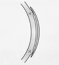
"Sunken graft" may not be the best term to describe this outcome. "Sunken" gives the impression that the graft-host junction drops off as if the grafted tissue lies recessed within the host tissue deeper than intended. We've never seen that happen. What we frequently see is an elevated bank at the graft-host junction that may or may not run the entire circumference of the graft. This bank occurs more frequently when running sutures, which may be single or double and may be used with interrupted sutures on the same graft, are present. In cases with only interrupted sutures, embankments are less common and less elevated when present.
Running sutures are typically removed about one year after surgery. Most banks decrease significantly or disappear completely soon thereafter. Until that happens, irregular topography of the graft often requires a rigid contact lens for adequate vision, especially if the fellow eye provides no or little help in vision. The presence of a bank at the graft-host junction makes this a tough job.
Determine Candidacy for Contact Lenses
If no complications occur following PK, most cornea specialists consider the eye ready for a contact lens about three months after surgery. Once that patient is in your chair, perform a thorough preliminary exam prior to trying on any contact lenses. A careful refraction with documentation of best-corrected visual acuity (BVA) with spectacles will determine if contact lenses should be considered at all. Generally, if BVA is 20/30 or better, we consider placing a contact lens on this potentially fragile surface unnecessary. If BVA of the grafted eye is 20/30 or better with spectacles and anisometropia was the reason for considering contact lenses, you may wish to fit the fellow eye with a balancing lens.
Corneal topographical mapping is essential for determining the need for contact lenses. The only PK cases we fit in contact lenses are those with irregular topography in the visual axis that causes BVA of 20/40 or worse.
Soft Lens Trial
The intent of a contact lens is to mask the irregularity of the graft. This makes one think initially of using a rigid lens. However, a bank can make that very difficult. We occasionally try a soft lens first because we expect less trouble getting it to fit over the rise and fall of the bank. Often these corneas are relatively flat and need a plus lens. In cases where the prescription is over +3.00, the thickness of any spherical lens can mask some of that irregularity. Trying a lens on the eye will show you how much will be masked. We have also had some success masking graft irregularity with the formerly Paragon, now X-Cel Flexlens Tricurve lens. Achieving centration and movement of soft lenses over a PK is usually not difficult. However, bubbles under the soft lenses are common when they cannot accommodate the slope of the bank's inner surface (Figure 1). Sometimes these bubbles spread over the visual axis or allow the lens to dislodge easily, making soft lenses a poor option. When it is possible to use hydrogel lenses, remember that neovascularization is a legitimate concern with thick soft lenses.

Figure 1. Flexlens Tricurve SCL. Retains bubbles at the base of inner slope of the bank. |

|
Spherical Rigid Lens Trial
For those who like to keep things simple, spherical RGPs might be worth a try. If the bank shows minimal elevation or covers less than half of the circumference of the graft, the displacement from the bank may be acceptable. In our experience, however, a spherical lens of typical diameter rarely works if a bank is present. It often centers over the steepest part of the bank with unacceptable decentration and bearing on the graft-host junction (Figure 2). In the unlikely event that an RGP centers over the graft, vaulting from one side of the embankment to the other will cause excessive clearance over the graft, heavy bearing on the bank and excessive edge standoff with bubble formation outside the bank (Figure 3). The high clearance over the graft can also allow bubble formation inside the bank (Figure 4). The SoftPerm lens can help center a rigid surface over the grafted cornea and usually allows less bubble formation peripherally. However, central bubbles remain common since the rigid lens still tends to vault the graft from bank to bank. Oxygen deprivation is a concern with the SoftPerm lens as well.

Figure 2. Spherical RGP centering over and bearing on the steep bank. |

|
Reverse Geometry Lens Trial
Reverse geometry lenses (RGLs) can achieve decent centration by matching the steep outer slope of the embankment. This also prevents excessive edge standoff and peripheral bubble formation. However, even when close alignment with the central cornea is achieved, the design of the RGL will not accommodate the inner slope of the bank. This occasionally allows bubbles to form inside the bank that will either disturb vision (if close to the visual axis) or make the lens easy to lose (Figure 5).

Figure 3. Spherical RGP, excessive edge lift outside the bank.
Small Diameter Custom Lens
There are so many problems with going over the bank that we often attempt to stay within its confines. The incision in the host tissue is usually about 7.5mm in diameter. If a bank is present, the inner slope will extend about 0.5mm into the grafted tissue. This leaves about 6.5mm of cornea that is usually not too difficult to fit. Figure 6 shows a spherical RGP measuring 6.0mm in diameter settling well within the inner slope of the bank. That leaves about 0.5mm of room for this lens to move on the corneal surface. Rapid flattening of peripheral curves helps prevent the edge of the lens from causing epithelial damage to the base of the inner slope of the bank. We very rarely find bubble formation with this type of lens, and it is usually successful masking the irregularities of the graft responsible for decreased vision. The most common problem with this small lens is dislocation when the inner slope of the bank is not steep enough to keep the lens from escaping. As long as this lens remains within the bank, the chance of neovascularization from hypoxia or mechanical irritation at the incision site are greatly decreased.

Figure 4. Spherical RGP with excessive clearance and retained bubbles. |

|
From Third Month to One Year
Once a PK patient is fitted with contact lenses, we follow up within a week, at one month and at least every two or three months thereafter. Monitor all PK patients in contact lenses for signs of neovascularization and mechanical irritation from excessive
bearing. With a small-diameter RGP, patients may experience lens dislocation after months of successful wear. This may happen if the embankment around the sunken graft decreases prior to the removal of the running sutures. At one year, when the running sutures are removed, the bank usually disappears.

Figure 5. Menicon Plateau RGL, better centration, less central clearance, but still tends to retain bubbles at inner slope of the bank. |

|
Little attention has been given to this type of cornea in contact lens literature. They are a difficult fit and the number of "banked" corneas encountered is relatively small. Also, the bank is a temporary feature. Putting off contact lens fitting until the running sutures come out is usually easier and sometimes appropriate. However, there will always be a few for whom contact lenses may be the only answer for functional vision during that nine-month period.

Figure 6. Custom 6.0mm diameter spherical RGP fitting within the bank. Parallel fluorescein pattern and no bubbles. Staining of the bank is due to excessive bearing by previous lens. |

|
The authors would like to thank their medical illustrator Justin Hanson and photographer Richard Hiatt.
To receive references via fax, call (800) 239-4684 and request document #67. (Have a fax number ready.)


