Instruments
Instruments and Technology
By John Mark Jackson, OD
July 2001
Sure, it's a cliché, but it's true: a picture really is worth a thousand words. How many times have you wished you had a way to photodocument a lid lesion to monitor growth over time or capture an image of a corneal ulcer to keep tabs on its resolution? Traditional solutions are quite costly: a digital photo slit lamp setup can cost many thousands of dollars beyond the cost of the slit lamp itself. Less expensive systems using film cameras have been available for years, but of course you have to wait for film processing. All in all, a convenient, easy and inexpensive solution has not been available to most practitioners.
|
|
|
|
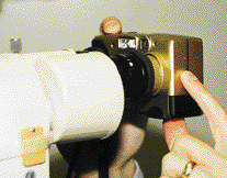
Figure 1. Camera lens shown held steadily against the slit lamp ocular. |
|
Go Digital
I have found a way to meet my documentation needs relatively inexpensively. I recently purchased a digital camera for home use, with no intention of taking it to work with me. On a hunch, I decided to try taking a picture of an RGP fit through the slit-lamp, and I was quite surprised by the quality of the result. I have used it over and over again and continue to find new uses for it as well as refine my technique for taking quality digital images.
Before I describe the technique I use for digital photography, I will discuss some essential digital camera features. Look for these features in a camera before you buy:
|
|
Digitize Your Contact Lens Practice |
|
Anterior and Posterior Segment Imaging Although there have been a number of digital imaging systems available in the past, with today's super-fast, large memory computers, advanced software, CCD cameras and improved monitors, they are better than ever. There are many advantages of digital imaging of the anterior segment or fundus with digital as opposed to 35mm or Polaroid imaging or drawing, including speed of capture and viewing and observation of multiple images. With modern, improved, easy-to-use software, excellent multiple images can be captured while you perform a slit lamp examination. You can take multiple images, dump the poor quality ones and store the good ones for future reference. You can send them to another practitioner for consultation. And perhaps best of all you can use them for patient education regarding lens deposits, corneal staining and neovascularization, just to name a few examples. One new ophthalmic image and capture management system is from Haag Streit (www.haag-streit.com) called EyeCap. It is used with the Haag Streit BQ slit lamp and has automatic archiving for ease of use. Security of patient data is considered in the software design. Improved RGP Lens Measurement Although digital gauges are not new, analog (dial gauge) microspherometers or radiuscopes can lead to dial reading and recording errors. The digital Contacto-Gauge made by Nietz and distributed by Polychem, Inc. USA (www.polychemusa.com) is one example of a modern radiuscope which records to the nearest 0.01mm for base curve measurement using digital technology. |
- The camera should have a small viewscreen on the back, like a small TV screen. This allows you to see directly through the camera lens, as you would with a single-lens reflex (SLR) camera.
- The camera should have manual flash and exposure settings to allow you to optimize the brightness of the images.
- The camera should have a high resolution rate, preferably 1.3 megapixels or above. The more megapixels, the more detail that can be captured, but cameras increase in price quickly as quality increases.
- The camera should have an AC adapter (or one that can be purchased separately) to power the camera. Digital cameras require lots of battery power, especially when using the viewscreen, and consequently drain batteries quickly. An AC adapter will save lots of money on battery replacement.
Here is the procedure I use to take pictures through the slit lamp:
- Turn on the camera, turn off the flash and make adjustments to exposure settings (experience will tell you if your photos are coming out too light or too dark). Turn on the camera's viewscreen.
- Focus the slit lamp on the object you wish to photograph (cornea, iris, contact lens, etc.) and lock the slit lamp into position.
- Hold the camera lens a few centimeters away from the slit lamp ocular. You should see the image of the ocular in the camera's viewscreen. Note the location of the ocular's exit pupil on the screen.
- Slowly move the camera towards the ocular, keeping the exit pupil as centered as possible. You'll see the image of the eye come into view. Make fine adjustments to the camera position as you hold the camera against the ocular to find the brightest, best-centered image.
- Once the camera is against the ocular and the image is in view, press the shutter button to capture the image. The photo should then appear on the viewscreen for a few seconds to verify that an image was captured.
- I usually take three or four images this way, refocusing the slit lamp and starting over again each time, and perhaps changing the exposure settings to get a brighter or dimmer image as needed.
Storing and Using Images
Once you have captured your image, you need to transfer it to your computer. The exact method will vary from camera to camera. Some cameras store images on a standard 3.5 inch floppy disk, but most will connect to a computer using a serial or USB (Universal Serial Bus) cable. I would strongly suggest purchasing a camera with a USB connection if you have a USB port on your computer, as transfer times are much faster than with a serial connection (seconds versus minutes). Refer to the camera's manual to determine the exact transfer method.
After the image has been downloaded, you can view it with software that comes with the camera. Most cameras come with some sort of simple photo-editing software package. Once you have your photo on your computer screen, evaluate it for sharpness, brightness and overall quality. Use these qualities to refine your technique. For example, if the photo appears washed out, decrease the brightness of the slit lamp beam or change the exposure setting on the camera to compensate. You can also adjust the brightness and contrast of the image using the editing software, but it is best if you start with a properly exposed photo. If the image is out of focus, you may have to experiment with the focusing of the slit lamp. Also, make sure that you are holding the camera steadily against the ocular as you take the photo.
|
|
|
|
Figure 2. Camera held several centimeters away from the ocular. Note the image of the ocular and the exit pupil in the viewscreen. |
|
You may choose to simply store your images on the computer, or you may wish to make prints of them to place in the patient's record. Even fairly inexpensive color inkjet printers these days are capable of reasonably good results. Photo-quality inkjet paper or actual glossy photo paper (available at most office supply stores) will be necessary to produce acceptable quality on most printers. The print quality depends strongly on the quality and resolution of the original image. Be sure to always have the camera set on its highest resolution available. Lower resolution cameras (below two megapixels) will not produce quality images above 5x7 inches; more expensive cameras with higher megapixels can produce good larger size prints. You must find a balance between quality and price to suit your particular needs and budget.
Above all, remember that digital images taken in this manner still can't compete with the quality of regular slit lamp slide or film camera prints. For example, it is difficult to capture very fine corneal details like striae or microcysts with this method. However, it is quite possible to get good images of more gross findings such as corneal abrasions or ulcers, contact lens fluorescein patterns, lid lesions, iris detail and the like. A hand-held digital camera can be a simple and reasonably inexpensive way to bring photodocumentation into your contact lens practice.

|

|
Figure 3. Camera held against the ocular, prepared to capture an image. Note the image of the eye in the viewscreen. |
Figure 4. Entire setup showing patient in position, slit lamp and camera in place and ready to capture image. |
Patient Education Animation |
||||||||
Through the years I've purchased several pieces of high-tech equipment, and I've recently discovered educational software for eyecare practitioners. This new software is called Eyemaginations, and it has high-quality 3-D computer graphic animations of the eye.
Software developer Steve Sopher, OD, and his three sons have produced patient- (and staff-) friendly 3-D animations delivered with oral explanations of eye disorders, refractive errors, spectacle lens designs, eye diseases and eye surgeries all provided instantly to patients in the exam room and/or dispensary. These full-screen animations are highly detailed and in color. Digital still images can be easily selected to highlight a patient's condition for further explanation. You can also draw on these still images using your on-screen cursor as a marking pen. The Eyemaginations software is easy to use. While your patient dilates or sits at the optical dispensary, you or your staff member can select the patient's condition(s) from the on-screen library and walk away. The patient sees a thorough description of his condition(s) from the Eyemaginations program. The current library offers many choices of animations. For example, you can show patients floaters, why their eyes must be dilated, how glaucoma affects the optic nerve and vision or a LASIK procedure. The newest animations include:
Patients tell me that they truly understand their condition (such as glaucoma or astigmatism) much better after seeing the program. Because better patient education leads to better compliance, I feel that this practice management tool provides outstanding patient education valuable to any practice. |


