discovering dry eye
Retro-illuminating the Tear Film: New Use of an Old Technique
BY CAROLYN BEGLEY, OD, MS, & NIKOLE
HIMEBAUGH, OD
March 2001
For many years, tear break-up has been clinically measured by instilling sodium fluorescein into the eye and watching for the appearance of the first dark spot or break in the tear film. Use of sodium fluorescein dye provides a
detailed picture of changes in the full thickness of the tear film over time, but instilling fluid and dye into the tear film may change its stability. In addition, the sodium fluorescein technique cannot be used to observe tear break-up over a contact lens.

|
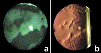
| Figure 1. Tear break-up with fluorescein (a) and retro-illumination (b). |

|
| Figure 2. Timed sequence of retro-illumination images. |
Go Retro
A technique with great clinical potential is retro-illumination of the tear film. For many years, retro-illumination has been used to observe cataracts. The light source of the slit lamp biomicroscope is aligned "on axis," and light is reflected off the retina through the pupil, resulting in a red reflex. Lens opacities will absorb the reflected light and appear dark or black in the red reflex. However, if the slit lamp optics are focused on the tear film rather than the crystalline lens, tear break-up can be observed. In this case, the image is caused by refraction of reflected light from the retina, rather than a shadow effect. Because the tear-to-air interface gives the tear film the greatest refracting power of the eye, small changes in tear film thickness during break-up cause large perturbations in the retro-illumination image. As Figure 1 illustrates, these changes correspond to tear break-up monitored using the traditional sodium fluorescein technique. The tear break-ups noted superiorly, centrally and inferiorly in Figure 1a are in the same location as the bright-dark bands in Figure 1b.
The same technique can be used to observe the progression of tear break-up in a patient wearing a soft contact lens. Figure 2 is a timed sequence of retro-illumination images from a soft contact lens wearer who kept her eyes open for 30 seconds. The retro-illumination image changes from very smooth (t=0s) to highly perturbed (t=30s).
The retro-illumination image results from refraction through the tear film, and thus is an optical technique that illustrates effects of tear break-up on vision. In Figure 2, much of the break-up is central, which would greatly affect vision. Tear film retro-illumination may have applicability in understanding the optics of tear break-up and also provides a high resolution map of tear film thickness changes with or without a contact lens. The pupil needs to be dilated to show a very large portion of the cornea. An infra-red light source can also be used, which would allow the pupil to dilate naturally.
Dr. Begley is an associate professor at the Indiana University School of Optometry and is also a member of the Graduate Faculty. Dr. Himebaugh is a graduate student at Indiana University School of Optometry, studying optical and visual effects of tear film break-up.


