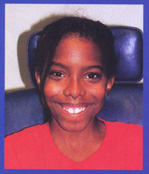
Controlling Strabismus with Contact Lenses
This case study explores how contact lenses can improve a patient's visual development and functioning.
By Darin Strako, OD, and Janice
Jurkus, OD, MBA
Contact lenses have been credited with providing better vision, more normal retinal image size, less distortion and a wider field of view than glasses. What is sometimes forgotten is that contact lenses can also help the patient's visual development, visual functioning and binocularity. Following is a case report that demonstrates the added benefits that prescribing contact lenses can provide.
|
|
|
|
Figure 1a and 1b. Patient exhibiting alternating
exo-deviation without correction. Notice pupillary reflex near the limbus of right eye when the left eye is fixating (Figure 1a) and near the limbus of the left eye when the right eye is fixating (Figure
1b). |
|
Patient History
An 11-year-old African American female, AS, was referred to the Illinois Eye Institute for an evaluation of her eye turn and to begin vision therapy. She also wanted new spectacles and contact lenses. Her ocular history revealed a large exotropia and high myopia diagnosed as an infant. She began wearing glasses at age 2 and contact lenses were initially prescribed for her at age 3. Our young patient's contact lens history included wearing RGP lenses, but due to comfort issues she preferred daily wear soft toric lenses. She was very happy with the vision and comfort of the soft toric lenses, but discontinued lens wear because one lens was torn. Prior to the lens damage, our patient wore her glasses and contact lenses an equal amount of time.
AS was born five weeks premature secondary to intrauterine growth retardation. Her birth weight was 2 lbs., 14 oz. She is small in stature compared to the average 11-year-old. Despite the early arrival, all developmental milestones were met, such as crawling by 10 months, talking by 12 months and walking by 14 months. Currently, she is in the sixth grade and is getting A's and B's in her studies. AS is healthy; no medications or allergies were reported.
Initial Exam
Our initial work-up confirmed her high myopia. Her glasses measured OD 17.501.50x161 and OS 20.25 2.50x005 with 20/30 distance acuity OU. Her near vision measured through her glasses was 20/60 OU. The cover test performed while wearing glasses revealed a constant alternating exotropia (CAXT) of 50 prism diopters at distance and 64 prism diopters at near. The near exotropia measured without correction was 55 prism diopters CAXT. Random dot stereopsis was not achieved with or without prism neutralization of 50 prism diopters BI. The Worth 4 Dot testing indicated crossed diplopia at distance and near with 50 prism diopters BI. Pupils, confrontation fields, color vision, EOMs were normal.
Anterior segment exam was unremarkable. Intraocular pressures measured with a non-contact tonometer were within normal limits. As expected, the dilated fundus exam revealed myopic crescents, stretching around the disks but neither eye had a staphyloma.
Keratometry measurements showed significant corneal cylinder, 47.75/50.75 @ 090 OD and 48.50/51.00 @ 090 OS. Our refraction slightly improved her visual acuity: OD 18.002.50 x165, 20/25 and OS 19.001.50 x015, 20/25.
Diagnosis
At this point our diagnosis was compound myopic astigmatism (CMA), constant alternating exotropia (CAXT) and myopic degeneration OU.
We were pleased and somewhat surprised that her aided acuity was good. The ability to see almost 20/20 stemmed from a few reasons. First, her initial correction was prescribed at age 2. This is within the sensitive period for vision development (up to age 8). Since she was fit with contact lenses at an early age, the improved retinal images enhanced her visual development despite her high myopia. Second, her alternating strabismus allowed her vision to develop equally because she alternates fixation eye to eye as opposed to constantly fixating with one eye. Finally, her fundus was not damaged from the thinning often seen in highly myopic eyes.
We assessed her binocular system. A cover test and stereopsis testing revealed a lack of bifoveal fixation. She had a constant alternating exotropia, which means she is unable to fixate with both eyes simultaneously. Random dot stereopsis was not achieved because it requires bifoveal fixation in order to see the image. Using relieving prism of 50 prism diopters BI to overcome her exotropia, we were unable to demonstrate stereopsis. We tested sensory fusion, the combining of retinal images within the visual cortex using a Worth 4 dot with 50 prism diopters BI relieving prism. This demonstrates the potential for sensory fusion at the patient's angle of deviation. AS was unable demonstrate sensory fusion.
Our plan was to prescribe contact lenses and reevaluate binocular functioning while wearing the lenses. If improvement was noted, vision therapy could more readily enhance visual functioning.
Contact Lens Fitting
When prescribing contact lenses for the high myope, it is critical to take vertex distance into consideration. Theoretically, the power at the corneal plane, determined by the vertex distance conversion, significantly reduces the amount of minus power.
Spectacle plane Corneal plane
18.002.50 x 165 14.751.75 x 165
19.001.50 x 015 16.501.00 x 015
We chose to prescribe soft lenses for AS because, in the past, she reported good vision and was comfortable wearing and handling this type of lens. RGP lenses were an option, but both the child and parent wished to continue using soft lenses if possible. Selecting a proper lens design is important. One of the considerations was the availability of lenses in high minus powers. Another was lens size. The diameter of the lens must be large enough to completely cover the cornea, but small enough to be easily applied to the small fissure width often found with children.
The refractive cylinder was less than 25 percent of the sphere power. Thus, we considered prescribing a spherical lens. Our first diagnostic lenses were spherical, the CIBA Vision D2LT (See Table 1).
|
TABLE 1: Diagnostic Lenses |
||
| OD | OS | |
| 8.3mm | Base Curve | 8.0mm |
| 14.00 | Power | 17.00 |
| 13.8 | AD | 13.8 |
| 1.251.25 x 165 | Sph/cyl OR | +1.501.50 x 015 |
| 20/20- | Visual Acuity | 20/25+ |
Our sphere/cylinder over-refraction improved both the quantity and quality of vision. Both lenses centered equally over the cornea and provided full corneal coverage. The median base curve moved smoothly measuring about 0.50mm to 0.75mm. The steeper base curve moved less and showed slight temporal vessel blanching.
By using a spherical diagnostic lens and a sphere/cylinder over-refraction, we were able to refine the refraction and improve visual acuity. We elected to order toric soft lenses; however, the CIBA soft toric is not available in a small diameter. Since a small diameter was desired, we ordered the CooperVision Hydrosoft Toric DW with a base curve of 8.2mm and a diameter of 14.2mm. Powers were OD 15.251.25x165 and OS 15.501.50x015.
Upon dispensing, vision through the lenses was 20/25+ OD, OS and OU. The fit showed lens movement of 0.5mm, complete corneal coverage and equal centration. Rotation measured less than five degrees on each eye. We were pleased with the lens performance, and we dispensed the Hydrosoft Toric DW for a two-week trial period. Upon returning for the two-week follow-up, we found no changes in fit, comfort or vision. Both the patient and her parents were very happy with the new lenses.
|
|
|
|
Figure 2. Patient showing ocular alignment when corrected with contact lenses. Pupillary reflexes are now centered and both eyes are
fixating. |
|
Binocular Function
We achieved our goal of improving acuity. Next, we evaluated binocular function. A visual efficiency evaluation with the new soft toric contact lenses revealed a significant difference in eye alignment. The cover test measured a 37 prism diopter intermittent alternating exotropia (IAXT) 95 percent of the time. This means that AS was able to fuse 5 percent of the time. This is the first time in the examination in which any fusion was elicited (Figure 2).
Functional vision with the lenses also improved. Stereopsis was achieved with the Random Dot E. She was able to note stereo vision in four of six test presentations. The Worth 4 Dot test resulted in sensory fusion at near (peripheral fusion) and alternating suppression at distance (central suppression).
The contact lenses have improved the patient's binocular vision in many ways. The reduction of the magnitude of the deviation from 64 prism diopters to 37 prism diopters at near is impressive. The magnitude of a strabismic deviation, when measured through minus spectacle lenses, is larger then the true angle of deviation. An exotrope, when wearing minus spectacle lenses, looks through BO prism. BO prism increases the magnitude of an exo-deviation. The dioptric power of a minus spectacle prescription incrementally increases the magnitude of the strabismic deviation. When contact lenses are used, the prism effects of the spectacle lenses are eliminated, revealing the true angle of deviation. Note that the opposite is true when using plus lenses. The measured angle is smaller through plus power spectacle lenses compared to the true angle of deviation.
The frequency of the patient's strabismic deviation changed from a constant alternating exotropia to an intermittent alternating exotropia. This means that AS has gone from fixating monocularly to being capable of fusion. Why is fusion present while wearing the contact lens? A large portion of the stimulus to fuse is elicited through the peripheral field of vision. It is far easier to fuse a large target that has peripheral cues than a small central target. For highly myopic patients, spectacle lenses create two different problems. First, minus lenses cause minification of the retinal image, which is more difficult to fuse than a larger image. Second, high minus lenses create a large amount of peripheral distortion. Distortion is perceived when looking obliquely away from the optical center. This leaves a small area of clear central vision in which to fuse. When contact lenses are used the amount of minification is decreased and peripheral distortions are eliminated. This gives the patient an environment that is much easier to fuse.
In summary, the contact lenses have made fusion possible. We were able to elicit both stereopsis (bifoveal fixation) and peripheral sensory fusion. These results make her an excellent candidate for vision therapy with a very good prognosis. With continued contact lens wear and compliance to a vision therapy program, it is possible to maintain ocular alignment and increase binocular function.
References are available upon request. To receive references via fax, call (800) 239-4684 and request document #80. (Have a fax number ready.)


