


SILICONE HYDROGEL COMPLICATIONS
Managing Silicone Hydrogel Complications
Identify and manage in-lammatory conditions and adverse events
associated with silicone hydrogel contact lens wear.
By Renée du Toit, Dip Optom,
MPhil, Deborah Sweeney, B Optom, PhD, Desmond Fonn, Dip Optom, MOptom, and Judith
Stern
Silicone hydrogels have been available worldwide for over three years. With the advent of this class of higher oxygen permeable materials, it was hypothesized that providing the cornea with sufficient oxygen during overnight wear would ensure little, if any, interference with the eye's defense systems. It was further postulated that the occurrence of microbial keratitis would be reduced if not eliminated, and ideally that adverse responses related to lens wear may also be eliminated or reduced. In a three-year clinical trial, Ren et al showed that all types of extended wear lenses suppressed surface epithelial cell shedding but that high Dk lenses did not appear to increase bacterial binding. This led the investigators to theorize that high Dk lenses may offer a significant advantage in safety for extended wear.
|
|
|
|
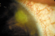
Figure 1. Contact lens peripheral
ulcer. |
|

Physiological changes due to hypoxia have been largely eliminated by silicone hydrogel lenses. There is virtually no corneal swelling and microcysts are rarely seen. Eyes look white due to reduced limbal hyperemia, and ghosting (emptying of the vessels) of neovascularization is seen in previous low Dk extended wearers.
However, it seems that even with adequate oxygen transmissibility, the eye's natural defense systems cannot always prevent infection and all types of inflammation. The presence of a contact lens when the eye is closed appears to place a burden on the defense systems, and adverse responses still occur. Although global surveillance of the events of microbial keratitis associated with the use of silicone hydrogels has revealed only about 14 cases to date, studies at Cornea and Contact Lens Research Unit (CCLRU)/ Cooperative Research Centre for Eye Research and Technology (CRCERT) and Centre for Contact Lens Research (CCLR) have shown that the rates of inflammatory conditions are similar to low Dk lenses, and some of the mechanically induced adverse events have higher rates.
Eyecare practitioners must be able to recognize the signs and symptoms and manage the various types of adverse events that manifest with contact lens wear. Accurate diagnosis will assist in identifying possible causes as well as influence the decision on management options. This can assist in advising patients about the likely time of abstinence from lens wear, chance of recurrence and strategies to reduce risks. While clear identification of an adverse event is essential, unfortunately widespread confusion still exists in the literature and in clinical practice. For example, the term "corneal infiltrate" is used to describe anything from a large infected ulcer to a few cells in the cornea. Infiltrates can occur in response to infection, inflammation and mechanical disturbance to the cornea.
|
|
|
|
Figure 2. Bulls-eye corneal scarring from a
CLPU. |

Adverse Responses
The signs, symptoms and management of adverse responses (AR) that are discussed below occur with all types and modes of contact lens wear; however, the emphasis will be on events specifically related to silicone hydrogel lenses.
Microbial or ulcerative keratitis (corneal infection) is the only serious event associated with contact lens wear. All other events can be classified as inflammatory or mechanically induced. The inflammatory events include contact lens peripheral ulcer (CLPU), contact lens acute red eye (CLARE), infiltrative keratitis (IK) and asymptomatic infiltrative keratitis (AIK). Mechanical events include contact lens papillary conjunctivitis (CLPC), superior epithelial arcuate lesion (SEAL) and corneal erosion. The standards of care vary considerably for each AR and across the spectrum of these ARs.
Corneal Infection
Microbial keratitis. This AR occurs very rarely. It is classified as a serious adverse event because vision loss may occur, although the reports of cases resulting in vision loss are extremely low. Significant delay or inappropriate treatment can affect the visual outcome. If a corneal ulcer is suspected to have an infective etiology, immediately institute aggressive treatment with antibiotics.
The presentation of MK can vary depending on the type and virulence of the micro-organism involved and the stage at which the patient presents. In general, excavation of the corneal epithelium occurs, exposing Bowman's layer and stroma, with marked necrosis and infiltration of the underlying tissue. The shape of the lesion is usually irregular and satellite lesions, smaller lesions adjacent to the primary site of infection, may be present. An anterior chamber reaction is often observed in the active stage. Patients presenting with MK are usually very symptomatic, and the condition worsens without intervention.
Prompt attention and treatment are mandatory because infection associated with virulent organisms can progress and cause severe corneal destruction within 24 hours. To establish whether the ulcer is infectious or not and the type of microbe involved, perform a corneal scrape and appropriate microbiological investigation. In addition, guidelines for when to culture include the following: if the defect is more central than 2mm from the limbus, more than 2 mm in diameter, more than 20 percent of corneal thickness, grade 2 or greater anterior chamber reaction.
The literature on treatment supports both monotherapy with fluoroquinolones, such as Ciloxan (A ciprofloxacin, Alcon) Ocuflox (ofloxacin, Allergan) 1gtt q 15 min x 6 hours/1 gtt q 30 min x 18 hours, or fortified multi-antibiotic therapy. Both have proven effective, yet resistance is possible with either treatment. Treatment varies according to the stage and severity of the condition. Once the microbe has been identified, modify the treatment based on the sensitivity of the organisms to particular antibiotics.
|
|
|
|
Figure 3. Contact lens acute red
eye. |
|

Inflammatory Events
Contact lens-induced peripheral ulcer CLPU (Figure 1) is a circular, well circumscribed, dense, yellowish-white, focal corneal infiltrate (0.2-2.0mm in diameter) with an overlying epithelial defect that stains with fluorescein, with diffuse infiltration of the surrounding stroma in its active stage. It is located in the peripheral to mid-peripheral cornea. CLPU is characterized by marked limbal and bulbar redness, usually localized to the quadrant adjacent to the lesion. Redness is the most common symptom reported by the patient, followed by pain or soreness, irritation or watering. CLPU is usually unilateral and typically features a single focal infiltrate; however, a number of infiltrates can occur, although this is rare. In severe cases, there may be mild anterior chamber involvement and photophobia. In mild cases, a patient may be asymptomatic and present to the clinic only with a scar as indication that an event occurred. CLPU can mimic early MK; however, CLPU symptoms are milder and begin to recede immediately on discontinuation of lens wear. The events usually resolve in a characteristic bulls-eye scar (Figure 2). The patients who present to our clinics with healed peripheral circular scars without treatment highlight the self-limiting nature of CLPU events.
Contact lens-induced acute red eye CLARE is classified as an inflammatory reaction following overnight sleep with contact lenses (Figure 3). It is usually unilateral. The patient typically presents with marked conjunctival redness and irritation or pain. Approximately one third of CLARE patients in our experience are woken by severe pain. Other symptoms noticed on waking or shortly after include discomfort and watering. The most characteristic feature of the condition is the presence of fine diffuse cellular infiltration of the peripheral to mid-peripheral cornea with clusters of small focal infiltrates interspersed or extending into the clear cornea. The conjunctival redness and infiltrates are circumferential in nature, the infiltrates appearing to "stream" from the limbal vessels. If infiltrates extend into the pupillary area, the patient may experience photophobia. The extent of involvement ranges from 10 to 360 degrees of the corneal circumference, although most commonly only 75 percent is affected. There is sometimes epithelial involvement, but if present, is limited to minimal punctate corneal staining over the infiltrates. The infiltrates are found in the anterior stroma. The posterior stroma and anterior chamber are usually unaffected.
Patients should discontinue lens wear until corneal infiltrates resolve. Ask patients to use non-preserved artificial tears and cold compresses during the acute phase of the condition. Frequently monitor patients until the infiltrates resolve. If there is any worsening of the event, institute therapeutic treatment immediately. Patients need to be counselled about the possibility of recurrence before resuming lens wear. The fact that infiltrates resolve on cessation of lens wear speaks to the relatively mild nature of the event.
|
|
|
|
Figure 4. Infiltrative
keratitis. |
|

Infiltrative keratitis IK (Figure 4) is a relatively mild event characterized by anterior stromal infiltration with or without epithelial involvement in the periphery to mid-periphery of the cornea. IK is usually accompanied by moderate redness and mild to moderate irritation. Photophobia may be a symptom if the infiltrates extend beyond the pupil margin. Lens wear should be discontinued until the condition resolves.
Asymptomatic infiltrative keratitis AIK resolves rapidly on discontinuation of lens wear. It features small focal infiltrates and/or mild diffuse infiltration. If the patient exhibits a small number of focal or only slight diffuse infiltrates without staining or redness, lens wear can be safely maintained. However, discontinuation of lens wear is usually recommended until the redness and/or staining has resolved, which usually takes a couple of days. Patients should be followed until the infiltrate resolves. Prophylactic antibiotic medication can be prescribed if any epithelial loss occurs in any of these inflammatory events.
Mechanical Events
Contact lens papillary conjunctivitis CLPC is a condition that largely affects the palpebral conjuctiva of the upper lid. The condition is characterized by enlarged papillae and redness. CLPC can be generalized, involving the whole of the tarsal conjunctiva (Figure 5), or localized (Figure 6), involving only a limited area in the central tarsus nearest the lid margin. Preliminary evidence indicates that patients wearing silicone hydrogel lenses are more disposed to localized CLPC than hydrogel lens wearers. The incidence of generalized CLPC is similar with silicone hydrogel CW to low Dk hydrogel EW. The time to occur varies considerably; with silicone hydrogel lens wear, the average is 11 months, ranging from six to 17 months.
|
|
|
| Figure 5. Generalized contact lens papillary conjunctivitis. | Figure 6. Localized contact lens papillary conjunctivitis. |


Generalized CLPC may cause moderate to severe patient symptoms. These include itching, irritation or lens awareness, a stringy or ropy discharge, excessive movement of the lens and blurred/fluctuating vision due to the movement or deposition on lenses. The symptoms in localized CLPC are generally much milder with slight irritation or foreign body sensation often the only symptom initially. If the condition advances, there is an increase in mucous production, which causes an increase in lens deposition. This in turn causes a decrease in visual clarity and an increase in lens movement and lens edge awareness. Itching may also be associated with CLPC, especially after lens removal.
With generalized CLPC, management options include frequent cleaning or replacement of lenses, a decrease in wearing time or a change in mode of wear (EW to DW), lens type or material. Mast cell inhibitors and/or steroids may be used to manage recurrent events. Tarsal redness decreases significantly but does not tend to return to baseline levels, and although papillae decrease significantly in size, these may remain dispersed over the entire tarsus. When lens wear is discontinued in localized CLPC, redness usually resolves in two to four weeks; however, the papillae, while smaller, tend to remain. Localized CLPC has a tendency to recur in approximately 50 percent of cases if silicone hydrogel CW is resumed. Current management strategy is to change to frequent replacement low Dk soft daily wear or daily disposable lens wear.
|
|
|
|
Figure 7. Superior epithelial arcuate
lesion. |

Superior epithelial arcuate lesions SEALs (Figure 7) present as a thin arcuate lesion in the superior cornea, with significant overlying staining and occasional underlying diffuse infiltrates. The edges of the lesion are often irregular and may be slightly roughened or thickened. Approximately one third are asymptomatic in silicone hydrogel EW, with the most common symptom being foreign body sensation or irritation. Approximately 40 percent have underlying infiltrates, and a third exhibit stromal glow of fluorescein. Time of occurrence of the first event varies widely; for example, in studies at the CCLRU, the time of onset has ranged from one to 20 months.
Discontinuation of lens wear until resolution of the SEAL, including infiltrates, is recommended. Resolution usually occurs within 24 to 48 hours, but may take one to two days longer if infiltrates are present. SEALs tend to recur in approximately 50 percent of cases, but the time to recur varies widely among patients. There is usually no scarring on resolution or after repeated events. Change the silicone hydrogel lens type and/or base curve especially with recurrence of SEALs, or discontinue silicone hydrogels in favor of a more flexible lower Dk soft lens for daily wear after repeated episodes of SEALs.
|
|
|
|
Figure 8. Corneal erosion. |
|
Corneal erosions Erosions (Figure 8) or abrasions can occur from mechanical trauma (fit, lens defect, trapped foreign body or on insertion and removal) or overwear. The damage is usually limited to layers anterior to Bowman's. The signs and symptoms can vary widely depending upon the cause. If the depth of the erosion is limited to the superficial one to three layers of the epithelium, the event is often asymptomatic; however, if it is deeper, moderate to severe pain, watering and blepharospasm may be present. If infiltrates are present, the event is classified as infiltrative keratitis, and is categorized as such with the appropriate management strategy followed.
If the erosion is small (<0.5mm) and superficial, lens wear should be discontinued for 12 to 24 hours, and the patient should be monitored. For large (>0.5mm) superficial or deep erosions, lens wear should be discontinued for at least 24 hours and not resumed until complete resolution. It is important not to patch the eye, as this significantly increases the risk of secondary infection. Prophylactic antibiotics may be used in severe cases; however, preservatives in these solutions can delay healing. Artificial tears can be used if discomfort is present.
Conclusion
With practitioner and patient education, all of these adverse events can be managed effectively. As with all contact lens practice, patients should be educated and regularly reminded that they should check every day whether their eyes look good, feel good and see well. Instruct patients to remove their lenses if they suspect a problem and immediate care should be available to any patient who suspects an adverse response. Patients should also be informed how to avoid risks with their contact lens wear. Most importantly, they should never wear a lens overnight if it is uncomfortable or if they are unwell. All contact lens patients should have a current pair of spectacles if they need to discontinue lens wear. With patient awareness and prompt and effective practitioner intervention, adverse responses can be managed with confidence.
Visit www.siliconehydrogels.org for more information.
Renée du Toit, Deborah Sweeney and Judith Stern are at the Cornea and Contact Lens Research Unit at the University of New South Wales in Sydney, Australia.
Desmond Fonn is at the Centre for Contact Lens Research, School of Optometry, at the University of Waterloo in Waterloo, Ontario, Canada.