contact lens case reports
Small Diameter GP Lenses
For Steep Nipple Cones
BY PATRICK J. CAROLINE, FCLSA, FAAO, & MARK P. ANDRÉ, FCLSA, FAAO
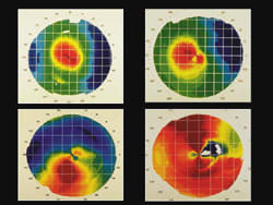
Keratoconic corneas may be topographically classified as nipple, temporal, oval and globus (Figure 1). The nipple form manifests as a central ectasia approximately 4.0mm to 5.0mm in diameter. The small central conical area is surrounded by 360 degrees of essentially "normal cornea." These corneas may be susceptible to apical touch with GP lenses, resulting in excessive corneal staining and scarring.

|

|
| Figure 1. The four types of keratoconus: upper left nipple, upper right temporal, lower left oval and lower right globus. | Figure 2. The patient's right eye with a central epithelial defect, swirl staining and stromal scarring. |
Case in Point
Patient JK presented to our office with a 33-year history of keratoconus. The patient recently began experiencing decreased wearing time and visual acuity in the right eye only. His contact lens corrected visual acuity OD was 20/100. His keratometric readings were greater than 60 diopters in both meridians with 4+ distortion. Parameters of his right lens were base curve 6.78mm (49.75 diopters), power 4.00D and diameter 9.0mm. Slit lamp examination showed the lens rocking on the corneal apex with significant horizontal decentration. Following lens removal, the right cornea exhibited a central epithelial defect, "swirl staining," and pronounced stromal scarring (Figure 2).
|
|
|
|
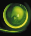
Figure 3. The optimum 8.3mm lens on the patient's right eye, with Ks in excess of 65.00
diopters. Note the epithelial defect at the time of dispensing. |
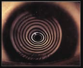
Figure 4. Post-fitting photokeratoscopy. Note the significant improvement in the central corneal distortion. |
A diagnostic fitting with a smaller diameter (8.3mm) Rose K lens revealed that a steeper 5.20mm (65.00 diopter) base curve was required to accommodate the central cornea (Figure 3). The final lens power was 18.50D with a resultant visual acuity of 20/60.
At the two-month follow-up, the patient reported improved comfort and wearing time with a visual acuity of 20/40. Photokeratoscopy photos revealed a mark-ed improvement in the central corneal distortion (Figure 4).
Patrick Caroline is an associate professor of optometry at Pacific University and an assistant professor of ophthalmology at the Oregon Health Sciences University.
Mark André is
director of contact lens services at the Oregon Health Sciences University.


