contact lens care
CL Solution Complications:
Know the Signs and Symptoms
BY JENNIFER L. SMYTHE, OD, MS, FAAO
Despite advancements in lens care systems, contact lens solution-related complications still occur. They are usually easy to resolve once identified, but differentiating between a solution-related response and other pathologic causes can prove challenging.
|
|
|
|
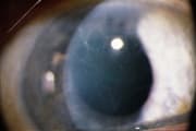
Figure 1. Corneal
infiltrates. |
Solution-Related Infiltrates
Solution-induced corneal infiltrates (Figure 1) commonly present bilaterally, are small (less than 0.5mm), diffuse and often appear in multiple numbers on the cornea. Corneal staining directly over these sterile infiltrates does not occur, but an associated diffuse superficial punctate keratitis is common.
Central infiltrates can reduce best corrected visual acuity and mild conjunctival hyperemia may develop. The presentation is similar to viral keratitis, so take a careful case history. Solution-related infiltrates respond quickly to treatment and recur if the patient returns to his habitual contact lens care solution.
Recognizing SLK
Although we rarely encounter thimerosal-preserved solutions that historically were strongly associated with contact lens superior limbic keratoconjunctivitis (SLK), practitioners have observed SLK with modern multipurpose regimens. Symptoms include mild burning, photophobia and decreased lens tolerance.
Slit lamp exam reveals hyperemia of the superior bulbar conjunctiva, superior corneal and limbal irregularity with punctate staining and hypertrophy of the superior tarsal conjunctiva, which are similar to SLK of Theodore. A history will help differentiate.
Keratitis or Non-Keratitis?
If a patient complains of dry, irritated eyes that result in reduced lens wear time and blurry vision, then suspect sensitivity to lens care ingredients. These individuals may display diffuse corneal staining in the inferior region of the cornea that may advance into the periphery, with the central cornea typically being the last area affected.
Patients who have non-keratitis exhibit symptoms identical to those who have keratitis minus corneal staining. Also, non-keratitis patients may have non-wetting of the ocular surface. Both presentations occur in healthy people who have normal tear production, but solution ingredients trigger an inflammatory response.
|
|
|
|
Figure 2. Pseudodendrites. |
|
Pseudodendritic Keratitis
Another viral "mimicking" lesion is the pseudodendrite (Figure 2). Solution-related lesions occur centrally or peripherally as a branching, twig or plaque-like epithelial or anterior stromal defect. Fluorescein staining is rare, corneal sensitivity is not affected and the branches do not have end bulbs.
Guilty Until Proven Innocent
To manage solution complications, regardless of presentation, discontinue the current care regimen and, upon resolution, change to a single-use lens modality, switch preservatives or dispense a preservative-free regimen.
Dr. Smythe is an associate professor of optometry at Pacific University and is in private group practice in Beaverton, Oregon.


