orthokeratology today
Using Tear Film Forces to
Correct ATR Astigmatism
BY JOHN MOUNTFORD, DIP. APP. SC, FAAO, FCLSA
|
|
|
|
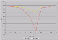
Figure 1. The difference in force in the steep (red line) and flat (yellow line) meridians. |
|
The force that works in the tear film under a reverse geometry lens is called the "squeeze film" force. The formula that describes it, Conway's formula, explains that the critical factor behind how the squeeze film force works is the difference between the apical clearance of the lens and the maximum tear layer depth at the edge of the back optic zone.
In essence, the lower the apical clearance, the greater the force and the greater the expected refractive change, and vice versa.
An Interesting Case
A patient who had a prescription of 1.00/-1.50 x 90 presented for ortho-k. Normally, I'd reject this case for two reasons: First, the cylinder is greater than the sphere, and second -- worst of all -- it's against-the-rule astigmatism. But, the topography maps looked interesting, showing apical radius (Ro) (flat) 7.41, eccentricity 0.34 and Ro (steep) 7.35, eccentricity 0.37. The sagittal depths were 1.5760 (flat) and 1.5874 (steep), which is about 11µm difference between the two meridians. That's about the sag difference required for a refractive change of 1.00D. So what would happen to this eye if I fit the steep meridian with the trial lens?
|
|
|
|
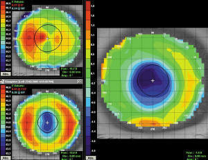
Figure 2. The subtractive map shows the change in astigmatism. Note the increased flattening of the steep meridian (horizontal) and the oval treatment zone in the lower left plot. |
In theory, less apical clearance would result for the steep meridian, providing a greater refractive change, and greater apical clearance would result for the flat meridian, providing a smaller refractive change. Figure 1 shows the difference in force between the steep and flat meridians. Note that the steep meridian (red line) has much greater squeeze force than does the flat meridian (yellow line).
A Fitting Success
As luck would have it, the trial lens proved almost identical to the final prescription lens, so I asked the patient to wear the trial lens for a week. The final refraction was +0.25/-0.50 x 90, with unaided VA of 20/15.
In the topography difference map (Figure 2), note the flattening of the horizontal meridian and the oval treatment zone. The cylinder measured at the 2.00mm chord was 0.50D x 90. Needless to say, both the patient and I were delighted with the result!
Fitting the steep meridian will work only if the sag difference between the two meridians is 15µm or less. Remember, the greater the apical clearance, the less the effect, and once the difference between the meridians exceeds 15µm, only about 1.00D of refractive change results.
Dr. Mountford is an optometrist in private practice specializing in advanced contact lenses for keratoconus, post refractive surgery and pediatric aphakia. He is a visiting contact lens lecturer to QUT and UNSW, Australia.


