corneal assessment
Corneal
Topography Basics and Clinical Applications
BY
MARJORIE J. RAH, OD, PHD
Advances in technology in the cornea and contact lens field have made corneal topography a key tool in many practices. The following is meant as a refresher of topography basics and to provide examples of various clinical applications of the technology.
|
|
|
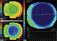
Figure 1. Refractive power difference map displaying the difference between pre- and post-PRK refractive surgery. |
Axial and Tangential Maps
An axial map shows an assessment of corneal curvature in diopters. Also called a sagittal or power map, it's commonly used to describe overall corneal shape.
A tangential map is a more sensitive map that you can use to detect more precise locations of a specific corneal defect. For example, when evaluating a patient who has keratoconus, the location of the cone's apex is more apparent when viewing the tangential map. When used with the difference display, a tangential map can also depict the change in corneal shape following treatment with orthokeratology or following a refractive surgical procedure.
Refractive Power Map
Axial and tangential maps measure corneal curvature, but not power. A refractive power map describes the refractive power of the cornea in diopters. This type of map is useful when evaluating the cornea following a refractive surgical procedure. Figure 1 shows a difference display of refractive power maps for a patient who underwent PRK surgery. The change in refractive power of the cornea is –4.50D, which corresponds to the manifest refraction change of the eye.
Elevation Map
|
|
|
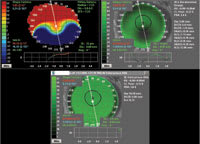
Figure 2. Elevation map and simulated fluorescein patterns of two lens designs for a patient who has keratoconus. |
An elevation map displays the difference in height of the corneal surface when compared to a best-fit spherical surface. You can use the information gleaned from this analysis to predict the fluorescein pattern when fitting GP contact lenses. Figure 2 shows the elevation map for a keratoconic cornea along with the simulated fluorescein patterns for two possible contact lens designs. The software allows you to make adjustments in the lens designs and to then view the resulting simulated fluorescein patterns.
Scratching the Surface
Each corneal topographer is different, but they all have extensive clinical applications. What I've discussed here are but a few brief examples of some of the basic applications of corneal topography. Many other features and benefits exist, but are too broad for the scope of this column.
Dr. Rah is an assistant professor at the New England College of Optometry where she works primarily in the Cornea and Contact Lens Service in patient care, teaching and research.


