readers'
forum
What
You Can Learn from the Tear Meniscus
BY
ETTY BITTON, OD, MSC, FAAO
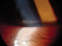
Examining the tear meniscus, also known as the tear lake or tear prism (Figure 1), can offer practitioners a "pool" of information. A thorough examination reveals information on the overall tear volume, the volume, the status of the lipid layer, the turnover rate of the tears and potential problems that may arise with contact lens wear.
Tear Volume
The tear meniscus height offers a subjective and indirect evaluation of the overall tear volume of the eye. A careful evaluation before you instill drops or dyes should reveal an inferior tear meniscus slightly larger than its superior counterpart. If this is not the case or an inferior meniscus is scant or absent, then you should suspect a preocular tear film (POTF) insufficiency. A normal tear meniscus height is between 0.2mm and 0.4mm for all age groups. A value of <0.1mm suggests a tear insufficiency. Patients who have a large palpebral aperture run the risk of further POTF evaporation.
For patients who are interested in lens wear, you need to perform other POTF volume tests such as the Schirmer or the Cotton Thread test to evaluate if the eye can sustain adequate contact lens wear. Counsel non-lens wearers to wear sunglasses when outdoors, especially during windy conditions, to limit further evaporation.
|
|
|
Figure 1. Slit lamp view of the tear meniscus. |
Meibomian Gland Secretion
A closer observation of the lid margin area will reveal a slight coloration (red and blue tones) within the meniscus and slightly above it. With every blink, sebum from the meibomian glands releases into the tear film. The coloration resembles a patch of oil in a puddle of water. Under higher magnification (16X to 20X), observe the meniscus area following a blink. The coloration should be slight and limited to the tear meniscus and the area slightly above it. If you observe the coloration over a greater surface of the cornea, then an excessive lipid layer is present. Although an adequate lipid layer is needed to limit POTF evaporation, an excess of sebaceous secretions makes for a very oily POTF, which becomes excessively sticky and attracts debris. Look for signs of "frothing" or "foaming" (small soap-like bubbles) at the outer canthus. If lens wear is possible in such patients, then recommend daily disposables to avoid reapplication of a soiled lens. If you fit a planned replacement lens, then instruct the patient to pay particular attention to cleaning the lens with a surfactant and an enzyme cleaner.
Absence of coloration can also identify a potential meibomian gland problem. In that case, perform expression of the meibomian gland by applying pressure to or squeezing the lid margin and observing the secretions. Yellowish to white paste-like secretions reveal meibomian gland disease and contribute to increased POTF evaporation because of an unstable lipid layer.
Tear Film Viscosity
You can observe bits of debris in the tear meniscus to determine how fast they move along the meniscus following a blink. How rapidly they move determines the viscosity of the POTF, which you can document as high, medium or low. Although this remains a subjective evaluation, high viscosity (slow movement of debris) reveals a thickened POTF; slower movement towards the punctum results in a slower POTF turn-over. Debris (from makeup or dust) and/or protein have an increased chance of adhering to lenses in these cases, so daily disposables are a good option to provide clean lenses everyday. In patients who have allergies, an increased allergen contact time can worsen symptoms. Artificial tears can provide beneficial relief by washing out the allergen from the ocular surface, especially in patients who have high-viscosity POTF.
Tear Flow
Observing the movement of debris or bubbles in the tear meniscus provides tear flow information. The movement should be in the nasal direction, toward the puncta, following each blink. A back-and-forth movement of debris or bubbles without overall nasal displacement can suggest a blockage of the naso-lacrimal route or an obstruction along the lid margin. Observe the entire length of the lid margin for scars, lesions, or mucus clumps that can obstruct the flow. A dilation and irrigation may be necessary to confirm the patency of the naso-lacrimal route.
Tear Clearance
Upon instilling fluorescein to color the tear meniscus, a prolonged tear clearance can suggest a poor tear turnover, an obstructed tear flow or incomplete blinking. This occurs more often in elderly patients afflicted with increased lid flaccidity, poor lid-globe apposition and ectropion with everted punctum. Reduced tear clearance can result in an accumulation of proinflammatory factors, such as cytokines, and can contribute to an inflammatory reaction of the ocular surface.
Look Closely, Save Time
A detailed observation of the tear meniscus and surrounding area provides information that can highlight potential problem areas — especially for contact lens candidates — and can help reduce chair time at future visits.
For references, visit www.clspectrum.com/references.asp and click on document #122.
Dr. Bitton is an associate professor of optometry at the École d'optométrie, Université de Montréal and is the Externship Director. She is also Secretary of the AOCLE.


