corneal
assessment
Photodocumentation
Without Going Broke
BY JOHN MARK JACKSON, OD, MS, FAAO
It's 5 pm on a Friday and a patient presents with a corneal ulcer. You'd like to have a photograph of it for patient education purposes and to monitor its resolution. You find yourself wishing you hadpurchased the slit lamp digital photography system that you saw at your last continuing education meeting, but boy was it expensive!
|
|
|
Figure 1. Resolved infiltrate scars photographed with this system. |
An Economical Alternative
The good news is that you can create your own digital photography system using your slit lamp without breaking the bank. The solution is simple: Use an inexpensive, consumer-grade digital camera to capture your photos. I first tried this a few years ago and have been very pleased by the low cost of the system and the high quality (Figure 1) of the results.
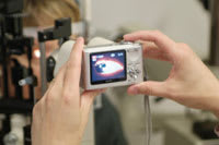
To capture a photo this way, simply hold the camera up to the slit lamp ocular (Figure 2). You'll be able to see the slit lamp image on the camera's LCD screen. Once you've centered the eye on the screen, gently depress the shutter button and you'll have a nice digital snapshot of your patient's eye.
Getting the Best Pictures
Here are a few tips to help maximize success with this method. Make sure that the camera lens is smaller than the ocular, which is true for most compact cameras. Choose a camera with as many megapixels as you can afford; this will allow you to zoom in on small details when viewing the image. The camera I use is a FujiFilm F10 with 6.3 megapixels. A large view screen on the camera helps.
Turn off the flash — the slit lamp is bright enough that you won't need it. Try different slit lamp magnification levels and different amounts of zoom on the camera to capture as much of the area of interest in the photo as possible. You'll have to practice a bit to get the best images, but it's well worth it.
Using Your Images
|
|
|
Figure 2. Hold the camera against the ocular to capture an image. |
Once you have the photos, you'll need to download them to your
computer and have a way to store and sort them. If you use a Macintosh, iPhoto is
an excellent way to store, sort and view images. If you use Windows, I recommend
a free program like Picasa (http://picasa.google.com/index
.html). Both programs
allow you to create folders that you can use to organize your photos by patient
name or ID number. You can choose to either store the images or to print them for
your patient record. Photo-quality inkjet printers have come down considerably in
price recently, and many of them can rival the quality of prints from a photo lab.
One last thought: Be sure to back up your photo library regularly!
Dr. Jackson is an assistant professor at Southern College of Optometry where he works in the Advanced Contact Lens Service, teaches courses in contact lenses and performs clinical research.


