contact
lens practice pearls
Case
Cracked by Corneal Topography
By Thomas G. Quinn, OD, MS, FAAO
|
|
| Figure 1. JB suffered from severe bilateral ptosis. |
Corneal topography is widely recognized as an invaluable tool in managing patients who have keratoconus and pellucid marginal degeneration as well as those undergoing refractive surgery and corneal molding. But it can also uncover other causes of visual problems that might otherwise go undetected. Here are two cases to illustrate this point.
Multiple Images for Many Years
Patient JB is a 53-year-old university professor who presented to the office sharing a history of perceiving multiple images in each eye since age 9. He also suffered from severe bilateral ptosis (Figure 1) and a right intermittent exotropia.
JB had seen numerous specialists over the years and no one had determined the cause of or provided relief from his visual disturbances. JB drew what he saw when viewing a spot of light from a laser pointer (Figure 2).
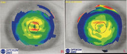
Best corrected visual acuity was 20/20 in each eye at 6M with a refraction of OD –1.75 –1.00 x140 and OS –1.00 –1.25 x066. Slit lamp examination of the anterior segment revealed no apparent abnormalities of the cornea or crystalline lens of either eye. The retina in each eye appeared quiet. Covering either eye didn't diminish the perceived multiple images. We then obtained corneal topographic maps (Figure 3).
|
|
|
Figure 2. JB drew what he saw when viewing a spot of light. |
Observing the topographic changes and their similarity to the images drawn by JB, we thought the visual disturbances might be associated with subtle topographic changes near the line of sight. We applied spherical GP lenses of the following parameters: OD 7.87mm base curve, 9.8mm/8.0mm overall diameter/optic zone, –2.00D; OS 7.80mm BC, 9.8mm/8.0mm OAD/OZ, –2.00D.
After allowing 10 minutes for settling, we assessed vision with GP lenses in place. Acuity of 20/20 was obtained with each eye. JB exclaimed,"This is quite remarkable! This is the first time I haven't seen multiple images in 44 years!"
JB indicated to me that, in addition to the visual improvement the contact lenses provided, the peace of mind he has gained from knowing why he has experienced the multiple images over these many years has been an even greater gift.
|
|
|
Figure 3. JB's corneal topographic maps. |
Breaking Open the Case
We had a suspicion that the observed topographical changes in this case were induced by pressure from the upper eyelids. Regardless, topography provided valuable information in uncovering the source of JB's visual difficulties and ultimately improving his visual well-being.
Dr. Quinn is in group practice in Athens, Ohio, is a diplomate of the Cornea and Contact Lens Section of the American Academy of Optometry and advisor to the GP Lens Institute.


