treatment
plan
Managing
Herpes Zoster Ophthalmicus, Part 2
BY
WILLIAM TOWNSEND, OD
Part 1 of this column (July 2006) described the case of a 37-year-old female who suffered from dermal and ocular complications of herpes zoster. In this column I'll focus on additional signs and symptoms as well as management strategies for this painful condition.
Nailing the Diagnosis
Herpes zoster ophthalmicus (HZO) typically begins with neuralgia of the lids followed up to a week later by the onset of dermal lesions. Hutchinson observed the association between HZV involvement of the nose and HZO, but ocular involvement can occur independently of nasal signs; one-third of patients who have HZO do not display Hutchinson's sign.
In contrast to herpes simplex, HZV involves subdermal tissue and thus may lead to scarring resulting in entropion, ectropion or trichiasis. Conjunctival hemorrhages and vesicles with follicular hypertrophy and localized adeno-pathy are common features of HZO. Nodular episcleritis and scleritis may present concurrently or months after the skin lesions have resolved.
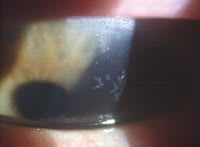
Keratitis occurs in over half of all cases of HZO; hypoesthesia is a common finding in these individuals. Corneal lesions may form a diffuse superficial punctate keratitis or a dendritic pattern that you could mistake for herpes simplex keratitis (Figure 1). HZO dendrites differ from HSK in lacking terminal bulbs, and wiping the surface of HZO lesions yields intact epithelium, whereas wiping HSK lesions leaves full thickness epithelial ulcers. Anterior stromal infiltrates occur in 40 percent of HZO cases.
|
|
|
Figure 1. Dendritic pattern of HZO corneal lesions. |
Management Strategies
Initiating systemic antiviral treatment within 72 hours after the onset of the skin lesions significantly reduces the probability of post-herpetic neuralgia (PHN).
Use of oral acyclovir prescribed at 800mg PO 5 times per day early in the course of the disease often reduces pain, duration of skin lesions and complications of episcleritis, scleritis and keratitis. Oral antivirals dramatically reduce the need for adjunctive steroid therapy. Newer antiviral agents famciclovir and valacyclovir exhibit better oral bioavailability and require less frequent administration, typically t.i.d. Concurrent use of oral steroids with antivirals may reduce dependence on pain medications and hasten healing of skin lesions.
Topical steroids are also useful in treating corneal, scleral and uveitic manifestations of the disease. Cycloplegic agents help minimize the discomfort and complications of corneal and uveal disease. Steroids do not exacerbate HZO keratitis as they do in herpes simplex keratitis. Taper steroids gradually, over a period of weeks or even months.
PHN is difficult to treat and typically has a clinical course of months or years. Skin lesions respond to Burrow's solutions, calamine lotions and creams. Topical capsaicin reduces pain once skin lesions have healed. Oral cimetidine can reduce itching and pain. Anticonvulsants such as gabapentin and carbamazine can effectively treat PHN. Tricyclic antidepressants and narcotic analgesics can minimize pain and discomfort.
You may find it useful to work with primary care providers and pain specialists in managing patients suffering from severe PHN.
Dr. Townsend is in private practice in Canyon, Texas, and is a consultant at the Amarillo VA Medical Center. E-mail him at drbill1@cox.net.


