treatment
plan
Caring
for Bullous Keratopathy
BY
WILLIAM L. MILLER, OD, PHD, FAAO
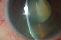
A benefit of working in a multi-doctor practice is the ability to see many interesting cases and to offer advice when needed. In one such case, the patient experienced pain in his eye with no light perception. He has a longstanding history of glaucoma with multiple medication and surgical therapies. A bulla encompassing nearly half of the cornea was evident (Figure 1). Diagnosis was extreme bullous keratopathy.
Signs and Symptoms
Bullous keratopathy (BK) is typically described by the underlying cause such as Fuchs' Dystrophy, aphakic/pseudophakic, post radial keratotomy, chronic highly elevated intraocular pressure and anterior uveitis. Each disease or condition alters the endothelial pump and barrier system to allow fluid to accumulate in the corneal stroma, which finds its way to the epithelium and produces bullae.
Typically bullae cause a great deal of pain and hinder functional vision. Microcystic edema may also be present as well as folds in the posterior limiting lamina.
|
|
|
Figure 1. This patient's bulla encompassed nearly half of his cornea. |
BK Treatment
Base treatment on the severity of the BK with the goal of decreasing corneal edema and discomfort. Initial treatments for mild to moderate cases of bullae include the use of bandage contact lenses, hyperosmotics and nonsteroidal anti-inflammatories (NSAIDs). NSAIDs can reduce the corneal pain, but use them with some caution because they may rarely cause corneal melting if the anterior corneal surface is severely compromised. Nevanac (Alcon) or Xibrom (Ista Pharmaceuticals) may be good newer choices for NSAID delivery with fewer side effects and lessened dosage regimens (t.i.d. for Nevanac and b.i.d. for Xibrom).
Hyperosmotics are fine for mild BK cases, but may be ineffective in severe cases. Knezovic et al (2006) has provided useful data to help identify patients for whom hyperosmotics such as 5% NaCl solution are helpful. Their research shows that in the early stage (stromal edema) of the disease, hyperosmotics can decrease corneal edema and improve visual acuity. Once epithelial edema with bullae develops, hyperosmotics are less effective. In reference to pachymetry, patients who have central pachymetry readings less than 613μm and peripheral readings less than 633μm are also suitable candidates for hyperosmotic therapy.
Contact lens choices for decreasing BK pain include silicone hydrogel lenses. Patients will likely need to wear them in an extended wear modality. I prefer Acuvue Oasys (Vistakon) because of its low modulus, although its use in this capacity is off-label.
Consider surgical intervention in cases of recalcitrant BK or subsequent severe scarring. Corneas scarred from frequent BK epi-sodes may need a penetrating keratoplasty. Newer surgeries such as deep lamellar endothelial keratoplasty and Descemet's membrane stripping are indicated when a patient suffers from Fuchs' dystrophy and has little to no corneal scarring.
Other treatment options include conjunctival flaps, anterior stromal puncture (ASP) and transplantation with amniotic membranes. ASP is for patients who aren't good candidates for a corneal graft. An amniotic membrane is reserved for patients who have little hope for visual recovery, but suffer from BK pain.
For references, please visit www.clspectrum.com/references.asp and click on document #130.
Dr. Miller is on the faculty at the University of Houston College of Optometry. He is a member of the American Optometric Association and serves on its Journal Review Board. You can reach him at wmiller@uh.edu.


