The
New Math of Corneal Staining
Hom, Milton M. OD, FAAO
The latest method for grading corneal staining may not always provide valid results.
We're all familiar with corneal staining. You wet the strip, instill fluorescein and observe using blue light and sometimes a yellow filter. You can look at the amount and location of staining, if at all present. You make a note and move on to the rest of the examination.
I used to think this was a relatively simple procedure. But if you scan the recent literature, you'll find that corneal staining has become the subject of a contact lens solutions controversy of numbers, grids and diagrams. Any practitioner with even a mild interest in contact lenses can't avoid the recent blitz of studies and statements. All of a sudden, this simple procedure has become incredibly complex with phenomenal nuances and sometimes unbelievable implications.
Causes and Consequences of Staining
Efron (2004) divides staining into six causes: mechanical, exposure, metabolic, toxic, allergic and infectious. Why is corneal staining important? The basic reason is that it forms a theoretical pathway to infection. A break in the epithelium is the first step for bacteria to get a foothold into the cornea. Jalbert et al (2006) demonstrated that large-area solution toxicity staining, covering four out of five corneal zones, is more likely to have corneal infiltrates.
On the other hand, the connection between infection and staining may be questionable. Willcox and Holden (2001) state, There needs to be damage to the epithelium in order for bacteria to initiate infection. The supporting reference from Klotz et al (1989) reports on an animal model of epithelial damage by filter paper.
Some of the recent literature omits the simple fact that corneal staining has a multitude of other causes and effects. Low-level corneal staining in itself doesn't mean there's a problem at hand. Efron (2004) and Guillon et al (1990) reported staining as high as 60 percent for contact lens wearers, but it's often low-level and clinically insignificant. As most fitters know, corneal staining can be normal. Several studies have found the mean grade for successful contact lens wearers to be 0.5 or less. Low-level staining of less than Grade 2 doesn't necessarily require action. For most patients, walking through life with Grade 1+ staining is a non-event rather than a crisis.
Clinical Significance and Grades
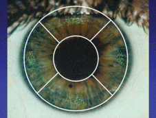
Researchers have used several grading systems to describe and quantify corneal staining. In the past, corneal staining was evaluated with 5 grades (0 to 4) over the entire cornea. Fortunately, systems have evolved. Most current classification systems divide the cornea into five zones: central, superior, inferior, nasal and temporal. Each zone is graded separately. The more sophisticated systems specify the number of punctate dots per zone. Other systems classify each zone with Grades 0 to 4 or something similar.
Despite their best intentions, clinical significance can sometimes be ambiguous. Let's consider some of the studies playing a role in this solutions war.
Grading Scales
One grading system garnering much attention is the Staining Grid, which divides the cornea into the typical five zones but grades on a percentage basis (0 percent, 10 percent, 20 percent, etc). The cut-offs for each percentage can be problematic. Let's say that less than 30 punctate or dots is 0 percent and 30 dots or more denotes 10 percent. In this case, 30 punctate is the cutoff between no staining and 10 percent staining. Here are some examples of how this method can be misleading.
Example 1: Zero Percent Grade
A dot cluster may be graded incorrectly if it falls between two sectors. In Figure 1, the 30+ dot cluster is considered 0 percent and 0 percent because each sector contains about 15 dots. I would think that we should consider the staining in this example as more than 0 percent and 0 percent.

|
Figure 1. If the dot cluster is greater than 30 punctate, but falls between two sectors, it would be considered 0 percent. |
Example 2: One Plus One Equals One
Along the same lines, two clusters may be almost equal in size, but one would be counted and the other would not. In Figure 2, the cluster for the left sector has 29 dots (graded as 0 percent) and the cluster in the right sector has 30 dots (graded as 10 percent).

|
Figure 2. Two clusters of equal size are shown. One would be counted, the other would not because it has less than 30 dots. |
Example 3: Is It Always the Solution?
Suppose the dots are located only in the inferior sector. Typically, staining from conditions such as dry eye or from distribution problems commonly occurs in the inferior sector. You would grade the staining in Figure 3 as 10 percent, but the cause could be something different from solution-related sensitivity. Also, in the previous Example 2, you may misinterpret three o'clock and nine o'clock staining as solution-related staining.

|
Figure 3. This cluster would be graded as 10 percent, but the cause could be something different from solution sensitivity. |
Example 4: Peripheral Dots
The dots in Figure 4 are located around the periphery, but number below the cutoff of 30 punctate for each sector. Under the Staining Grid system you'd grade the staining as 0 percent for all sectors. However, many solution sensitivity types of staining are located around the periphery.

|
Figure 4. The peripheral staining in this example would be graded as 0 percent under the grading system discussed. |
Statistical Significance
Researchers perform statistical tests to determine if a finding is actually significant. P-values equal to or less than 0.05 are considered statistically significant. In biostatistics, p-values are needed to validate a claim. You may see very large percentages, but if the readings have a wide variability and range (large standard deviations), it may not have statistical significance.
In the Staining Grid poster presentation (Andrasko, 2006), no p-values accompany the percentages. Although the percentages may be large in some combinations, a p-value is necessary to determine significance. Hopefully we'll see this information in the future.
Karpecki (2006) identified another example in which we could question statistical significance. A recent study suggests that ReNu MultiPlus (Bausch & Lomb) was associated with significant reduction in relative corneal sensitivity compared to Opti-Free Express (Alcon). They enrolled only four patients within each arm of the study, making an extremely small sample. Another confounding variable arises from the researchers' decision to include outliers within the final data set. One of the aesthesiometry readings in this study was significantly lower (20.00) than that of all the other data in either group. From a clinician's perspective, if you have a reading of 20 in one case when all the other measurements averaged 82.86 (with 100 percent representing the highest degree of comfort), you could come to the obvious conclusion that the patient registering a 20 would have trouble wearing contact lenses to begin with. As a result, most researchers would classify this particular data point as an outlier that they should footnote, but exclude from the final study results. If they had followed those scientific steps, the result would likely have shown no statistical difference between the solutions.
Other studies show quite the opposite staining response. In one open-label, multicenter study, researchers switched satisfied hydrogel (615 subjects) and silicone hydrogel (19 subjects) contact lens wearers from their habitual lens care product to ReNu MultiPlus. They recruited current users of Opti-Free Express or Complete (Advanced Medical Optics) multipurpose solutions. Graded slit lamp evaluation showed fewer statistically significant (P<0.05) occurrences of epithelial edema, epithelial microcysts, corneal staining, limbal injection, bulbar injection, tarsal conjunctival abnormalities, neovascularization and infiltrates associated with the use of ReNu MultiPlus compared with the subjects' habitual solutions (Figure 5). Since the introduction of the original ReNu MPS formula, the incidence of infectious keratitis, as reported by Poggio et al in 1989 and Cheng et al in 1999 hasn't increased. Over the past almost 20 years during which PHMB solutions in general have become the principal choice of tens of millions of contact lens wearers worldwide, a recent PubMed search reveals that no increase in the incidence of contact lens-related microbial keratitis has occurred.

Figure 5. Results from a study
comparing ReNu MultiPlus to Opti-Free Express and Complete.
Consider Results Carefully
Unfortunately, some of the conclusions from the aforementioned studies may bring about unwarranted fear about corneal staining for patients. History shows that medical scares are nothing new (cell-phone-induced brain cancer, acrylamide in potato chips and Alar in our food supply). Hopefully the new math won't add corneal staining to that list.


