Imaging in Cornea and Contact Lens Practice
Anterior segment photography isn't a glamorous tool. But it is an excellent complement to contact lens and anterior segment care and management. It's something that you can and perhaps should apply to most if not all of your patients.
|
|

Figure 2. A photograph that illustrates the fit of a GPlens. |
What's better than written notes or drawings? What says a thousand words in an instant? It's the photograph. With one quick look, you can tell that the eye in Figure 1 has an ocular surface problem, or what kind of fit a GP lens has in Figure 2.
A photograph is valuable because of the amount of information that an image can hold. When you read a patient's chart a year later, do you still understand its meaning? You may make detailed notes and even drawings, but even if these notes are understandable to you, they may not be clear to others.
Now look at those same notes with an accompanying photograph. Why can't all charts be so easy to read?
Here I'll explain how you can get started with anterior segment photography.
Choosing Your Camera
Is anterior segment photography expensive or difficult? It doesn't have to be. It's as simple as using a digital point-and-shoot camera. Sure, you can spend thousands of dollars more, but you don't have to. I use a five-year-old 3.34 mega pixel camera to take full-head pictures. I see many trauma patients from the emergency room, and it's great to be able to photo document facial as well as periocular injuries or pathology.
It wasn't too long ago that film ruled the world of photography. Film still offers better color fidelity and sharpness. But what we lose in those two attributes with digital photography we gain in convenience. It's heartening to know that you have a good image while the patient is still in the office rather than worrying about whether you got a usable image while waiting for the film to return from the laboratory.
Consumer digital cameras are capable of taking clinically useful photographs of the upper face, lids and globe. Such pictures are ideal for establishing landmarks like right and left eyes or lid position.
I photographed the patient in Figure 3 using the camera and its built-in flash at a distance of 2 feet with the lens set at a slight telephoto effect (to increase camera-to-subject distance).

Figure 3. Patient was photographed with just the camera and its built-in flash at a distance of 2 feet and the lens set at a slight telephoto effect. |

Figure 4. Photo can depict a frames position on a face, and may serve as a rationale for pediatric contact lens wear. |
Special Features
Today's consumer-style digital cameras may not have interchangeable lenses. Don't worry. You won't miss this feature because most digital cameras come with modest zoom lenses that work for most situations, if not for all. Fixed lens cameras also tend to be smaller. In fact, they're small enough to fit into a jacket or clinic coat pocket. Small also means that the camera is always nearby. Many a good photographic opportunity has been lost because a larger camera is usually stored away from arm's reach or is too bothersome to find and retrieve.
You've also heard about the mega pixel (MP). This is the feature that most buyers and users pay the most attention to. The inevitable question is how many mega pixels are enough. The good news is that even 3.0 MP is sufficient for most eye needs. Think of 8 MP or 10 MP only when you're trying to print a large poster.
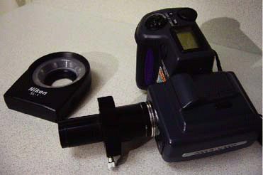
Most of the photographs I take are with a 3.34 MP fixed lens point-and-shoot camera. It has a macro capability and on-board flash for most full face photos, or of the forehead and eyes. For even closer shots of the eye itself, I added a front-threaded LED ring light. The ring light illuminates the eye better because at distances closer than 6 inches, an off-axis (of the camera lens) on-board flash cannot shed enough light. An on-axis light source (one that is positioned nearer to the camera lens) can (Figure 5).).

Figure 5. This photo of a subconjunctival hemorrhage was taken with an on-axis light source. |
The Right Light
When I see something interesting on the cornea, conjunctiva, irides or crystalline lenses, I use the camera with the slit lamp. I either hold my camera against the stationary ocular of the slit lamp or replace the ocular with an adapter tube and slide both the adapter and the camera into the space previously occupied by the ocular (Figure 6). The former approach is a bit more tricky because I have to line up the entrance pupil of the camera lens to the exit pupil of the ocular to ensure a full field image. With either approach, you often need a cooperative patient and sometimes a third helping hand (Figure 7).).
Taking great pictures of the eye isn't difficult or complicated. The critical success factors are appropriate lighting and a steady hand. In a full-face picture, the camera's own flash will usually freeze the minute movements that are inevitable. Some camera flashes, though, may highlight lightly complexioned faces too much, thus washing out the details of the face.
Sometimes the flash can wash out a particularly congested or hyperemic bulbar conjunctiva. It's dismaying to see how red an eye looks in the office, but it only looks one-third as red in the image. If this happens, stepping back a foot or more might reduce this reflection. Sometimes a bright room light will further enhance the light of the picture.

|

|
|
| Figure 6. Lens tube adapter for the ocular. |
|
Figure
7. This image of an amiadurone swirl was taken |
On a slit lamp, the illumination is more complicated. The brighter the light, the faster the shutter speed, which means that camera movement is minimized. But if there's too much light, corneal reflections and patient discomfort are more likely to occur. Use too little light and the picture becomes fuzzy because of camera shake or patient eye movements. A good picture deserves a balance of all of these factors.
Sometimes it's not the amount of light that matters but the quality of the light. Some slit lamp camera vendors offer fiber optic fill in illumination. By highlighting the surrounding tissues, the picture may actually be easier to see.
A Practical Approach
Let's get back to the practical. Following is a quick step-by-step approach to help you incorporate photography into your practice.
-
Have the camera ready and nearby. I hate to fumble for a camera especially if a patient is a bit anxious already. It also conveys confidence that you've done this before.
-
Sometimes, I take an overall head shot to get a frame of reference for the more magnified image. Or, I'll photograph some part of the medical record to ensure that I can match the image with the patient's chart later. Some practitioners even write down the exposure numbers and reference them with a patient name.
-
Determine the value expected of the photo. Is it a general photograph or is it positioned for risk management? Expect to handle either differently. The former requires fewer images and the latter more. I routinely take photos of a corneal foreign body and rust ring both before and after debridement, one to three days after initial treatment and at the time of discharge.
-
Particular to contact lenses is the relative appearance of the lens in situ prior to any vital stains or examination. What makes this valuable is the potential for patient education. Seeing this before any doctor intervention can be helpful toward better compliance.
-
If you're using this photographic image within the office and it's part of the examination procedure, then you don't necessarily need a model release. If you want to use the photograph but have no informative or identifying information on the image, then again, no specific model release is needed.
-
Once I've finished examining patients and taking photographs for the day, I remove the memory card and insert it into a memory card reader on my desktop computer. I have also transferred files between the camera and the computer using a special data cable. Either works well. Doing this daily translates to better characterization or sorting of the images.
-
I tend to back up my images by leaving them on the memory card and buying new cards as they fill up. I also have the images stored on a desktop computer, which is also backed up.
-
A word of caution about memory cards. They're not completely trouble free. If you happen to turn on the camera and then open the battery compartment and disconnect the battery, you will likely lose all the images on that memory card. This is a permanent error.
My use of photography during the course of a clinical day is mainly driven by curiosity, patient education and the goal of better patient care. All of these factors must somehow work together to minimize interference of the workflow of a clinical encounter. This isn't always possible! If photography ever does become a disruption in your practice, you must decide whether the value of the photo supersedes workflow. That isn't necessarily easy to determine either. Yet, I think the treating practitioner is the final arbiter as to what's appropriate.
Reimbursement for Photographs
Now you're probably thinking, can I ever get paid for taking these photographs? This depends on the reason for the photograph.
If a photograph is for patient education, then it's not likely to be billable to a medical plan. When it is billable, John Rumpakis, OD, of Lake Oswego, Oregon, an authority on third-party billing for optometrists, advises that photography can be reimbursable if the photograph is a medical necessity.
Dr. Rumpakis asks each practitioner to consider the relevance of the photograph to treatment and whether without it, the outcome would have been different.
Clearly, though, most practitioners such as myself like taking anterior segment photographs for better patient education.
For contact lens practitioners, it's an excellent way to demonstrate a particular risk factor. For instance, I would use a photograph to show the location of a particular corneal foreign body. In this form, I would bill the photo directly to the patient. Dr. Rumpakis advises also that this charge should be equal to what you charge a medical plan.
Improved Patient Management
Photography of the anterior segment can go a long way in helping your patients better understand their contact lenses, the nature of their care and the potential for complications.
Photography is more than just showing the condition. The value of photography is in its ability to present a tremendous amount of information to someone in a short span of time.
Patients understand and remember more from a picture than what we say. What someone may partially remember in words he will remember indelibly by imagery. Photography can even level the playing field with other practices that may offer services in other areas. Optometrist Michael Murphy's Swansea, Illinois, practice is less than a year old, and his use of anterior segment photography has definitely been a practice builder.
In summary, you can incorporate anterior segment photography into your practice simply and easily. You can do it quite frugally and yet yield valuable and relevant results, leading to better patient management.
For those who find photography rewarding and interesting, an investment in a more comprehensive system will only add value to your present practice.
Dr. Hom is Clinical Coordinator, Primary Care Optometry at the Department of Ophthalmology and Ron Robinson Senior Care Center of the San Mate Medical Center in San Mateo CA. He has written and lectured about cultural diversity, medical issues in eye care and high technology. You can reach him via his Web site at http://www.geocities.com/rchom/


