


Online Photo Diagnosis
By Gregory W. DeNaeyer, OD
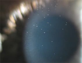
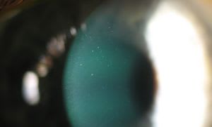
These photographs show mucin ball formation secondary to contact lens wear. The patient had been wearing his –1.50D OD and –1.75D OS Ciba Vision Night & Day lenses on an extended wear basis. He reported removing the lenses and sleeping without them once or twice a week. The patient replaced his lenses on a monthly basis. His visual acuity was 20/20 in both eyes, and he was asymptomatic. Mucin balls are thought to consist of collapsed mucin that results from mechanical interaction of a contact lens with the tear layer and corneal epithelium1,2. Mucin balls can indent the epithelium and pool fluorescein3. They should be differentiated from corneal microcysts, which are 10-micron to 50-micron vesicles that result from corneal hypoxia4. Mucin balls usually have no clinical consequence; in fact, they may be associated with decreased incidence of corneal inflammatory events5.
- Miller TJ, Papas EB, Ozkan J, et al. Clinical appearance and microscopic analysis of mucin balls associated with contact lens wear. Cornea 2003 Nov;22(8):740-5.
- Tan J, Keay L, Jalbert I, Naduvilath TJ, et al. Mucin balls with wear of conventional and silicone hydrogel contact lenses. Optom Vis Sci. 2003 Apr;80(4):291-7.
- Papas EB. Clinical significance of mucin balls.
http://www.siliconehydrogels.org/editorials/previous_editorial_eric_papas.asp accessed June 3, 2013. - Keay L, Jalber I, Sweeney DF, Holden BA. Microcysts: clinical significance and differential diagnosis. Optometry 2001 Jul;72(7):452-60.
- Szczotka-Flynn L, Benetz BA, Lass J, et al. The association between mucin balls and corneal infiltrative events during extended contact lens wear. Cornea 2011 May;30(5):535-42.