Online Photo Diagnosis
By Gregory W. DeNaeyer, OD, FAAO
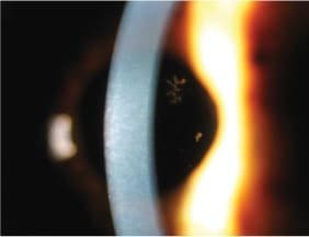
This picture shows a pseudodendrite in a 71-year-old patient who presented with Herpes Zoster Ophthalmicus (HZO).
Approximately 65 percent of patients who have HZO will have corneal involvement1,2. Herpes Zoster can affect the cornea in a number of ways including direct viral infection, antigen-anibody reactions, vasculitis, and neurotrophic keratitis2. Pseudodentrites that form are raised, plaque-like lesions of swollen epithelial cells that are without ulceration and are thought to result directly from the virus2. These of course need to be differentiated from dendrites that result from herpes simplex that are ulcerated and have terminal bulbs. Management of pseudodendrites involves only observation and palliative treatment with lubrication.

This patient was taking Famvir 500mg t.i.d. and doing well otherwise. For his cornea he was prescribed preservative-free artificial tears and Erythromycin ointment q.i.d. His pseudodendrite cleared in one month's time.
Reference
1. Liesegang TG. Corneal complications from herpes zoster ophthalmicus. Ophthalmology. 1985 Mar;92(3):316-24.
2. Kaufman HE, Barron, BA, McDonald, MB. The Cornea 2nd Edition. 1998 Butterworth-Heinemann 283-288.


