Online Photo Diagnosis
By William Townsend, OD, FAAO
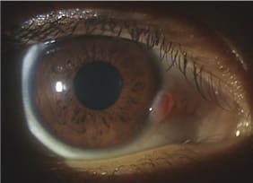
This 15-year-old male patient presented with a history of a "red spot" that appeared on the limbus of his right eye 18 months earlier. He noted that it became more injected at times. It had remained fairly consistent in size until several months ago when it began to grow. The patient could not remember any specific trauma to the right eye. His uncorrected presenting acuities were 20/20 OU.

Biomicroscopic examination revealed an elevated vascularized lesion straddling the right nasal limbus. The left nasal limbus had a much less developed and smaller lesion (second figure). The right eye mass was mobile, did not seem to be adherent to underlying sclera, and appeared to be non-pigmented. Fundus examination was entirely unremarkable. Our differential list included carcinoma in situ, amelanotic melanoma, scarring secondary to trauma, and an atypical pterygium. The very early age of onset was of great concern to us.

We arranged for the patient to be seen for biopsy/consult with an ophthalmologist. The ophthalmologist's initial findings concurred with ours, and after excising the mass he used cryotherapy to destroy any residual cells should the mass prove to be maliginant. Fortunately, the pathology report indicated that it was a benign growth exhibiting chronic inflammation possibly as a result of focal trauma.
The rapid appearance of any mass on the conjunctiva or cornea is cause for concern, especially in the young, and warrants a biopsy to rule out malignancy. Our patient was fortunate; in our clinic we have seen lesions with a similar appearance that proved to be malignant.


