January 2013 Online Photo Diagnosis
By Gregory W. DeNaeyer, OD, FAAO

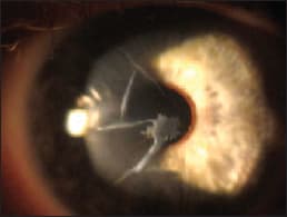
Figure 1 shows the right cornea of a 62-year-old patient who underwent bilateral radial keratotomy surgery in 1994. The patient complained of decreasing vision in both eyes since his surgery and that vision in his right eye was not improved with glasses. He reported failing in corneal GP and in hybrid contact lenses. Spectacle correction was OD +4.50 -3.00 X 098 20/200 and OS +6.00 -2.50 X 005 20/20. Slit lamp exam revealed thickened RK incisions in both eyes, with paracentral fibrosis of the right cornea. Corneal maps from topography showed severely oblate corneas OD and OS. The right topography had moderate irregularity (Figure 2).

The patient was successfully fit with scleral GP lenses with diameters of 18mm in both eyes, achieving visual acuity of 20/40 OD (Figure 3) and 20/20 OS.


