Our patient is a 43-year-old female who has extensive ocular history. She was born with congenital cataracts and nystagmus that were not discovered until the age of three months. Bilateral cataract surgery was performed, and her postoperative binocular aphakia was managed with both spectacle lenses and contact lenses. In 2006, she underwent intraocular lens implantation to her right eye only. Postoperatively, she had significant endothelial dysfunction and underwent an endothelial transplant (Descemet’s stripping endothelial keratoplasty [DSEK] procedure). The right eye was further complicated by high postoperative pressures that necessitated a posterior chamber tube shunt.
Today, her many ocular conditions are stable; however, she continues to have moderate-to-severe dry eye symptoms in both eyes. Her entering best-corrected visual acuities were right eye 20/200 and left eye 20/60. An external exam showed the eyes to be microphthalmic with a horizontal visible iris diameter (HVID) of approximately 10.2mm.
Deciding on the Right Lens
The patient had attempted 16.5mm scleral lenses approximately one year ago, but abandoned the lenses due to significant symptoms of discomfort. We diagnostically fitted the patient with 14.0mm diagnostic lenses.
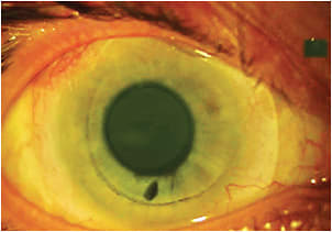
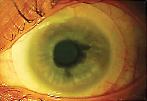
The lenses were a design that we had experimented with a number of years ago related to scleral lens wear for “normal” eyes. Our experiments with the 14.0mm lens design failed to provide the appropriate fitting characteristics on normal (11.5mm to 12.2mm HVID) eyes, and the project was abandoned. However, that was not the case for this patient’s microphthalmic eyes. The 14.0mm lenses showed an optimum amount of corneal and limbal clearance (Figures 1 and 2).


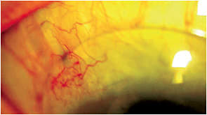
Fluorescein was placed onto the anterior lens surface to check for any peripheral areas of fluorescein leakage beneath the lenses. None was found, which indicated the presence of minimal or no scleral toricity. Subjectively, the patient was extremely comfortable with smaller-diameter scleral lenses. On the right eye, the 14.0mm diagnostic lens showed minimal interaction with the superior shunt and bleb (Figure 3), and we felt comfortable (at least for now) ordering the 14.0mm design.

The following lenses were ordered: OD 8.80mm base curve (BC), 14.0mm diameter, +0.50D power and OS 8.80mm BC, 14.0mm diameter, +25.00D power. To better manage the anticipated corneal edema, we ordered them in the highest-Dk lens material and with minimal post-tear lens vault and minimal lens thickness.
We will dispense the lenses to the patient and closely monitor her dry eye symptoms, her intraocular pressure, and her corneal and conjunctival response to the scleral lenses.
This case report illustrates the benefit of “hoarding” previously failed lenses, because you never know when the parameters and design of that particular lens may prove to be the perfect diagnostic lenses for a patient. CLS


