GP Lens Fitting in Keratoconus with Pterygium
This 31-year-old flight attendant from Cabo Verde, Africa has keratoconus with type II pterygium in both eyes. Recently, she visited our clinic in Brazil. She found us on the internet in her search for a better outcome, as she was not satisfied with her previous GP lenses that were fit in Portugal. These lenses exhibited severe apical touch OD, and she was not able to wear the OS lens because it would fall off of her eye.
When keratoconus is accompanied by another condition such as pterygium, it can be difficult to achieve the best possible contact lens fit. A pterygium may advance over the cornea until it reaches the area over the optical zone, negatively affecting visual acuity, or it may stop in any phase of its evolution.1 The latter appears to be the case for this patient, as she related that she has had it for a few years, and it had not advanced since it was noted entering the peripheral cornea.
Figure 2 shows the pterygium, with the neoformed fibrovascular tissue in a trapezoidal2 shape, entering the cornea about 2mm to 3mm (type II). Pterygium growth over the cornea may cause symptoms such as foreign body sensation, ocular burning, irritation, and excessive tearing; in healthy corneas, it may induce corneal astigmatism.3 Surgical removal of a pterygium can also change the anterior surface topography. It is furthermore quite common to observe recidivation of the condition. Yearly follow up is a good rule of thumb.

Management of the Case
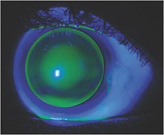
This patient has moderate keratoconus OD and advanced keratoconus OS (Figures 3 and 4). We instructed her to suspend lens wear for three days and then proceeded with lens refitting. The ectasia is centrally located, which in this particular case was helpful as it allowed us to fit a 9.0mm corneal GP lens that is positioned centrally and exhibits minimal movement, optimal tear exchange, no touch at the apex, and no interaction with, nor irritation of, the pterygium. The GP lens parameters were OD 54.50 x 45D base curves, 9.0mm overall diameter (OAD), 6.2mm optical zone diameter (OZD), –11.00D, visual acuity (VA) 20/20+3 and OS 64.00 x 45.00D base curves, 9.0mm OAD, 6.2mm OZD, –24.50D, VA 20/20–1


She received her lenses in two days and experienced nearly instantaneous adaptation, even in the left eye in which she had not worn a GP lens for nearly three months. Figures 5 and 6 show the left eye fit with this custom keratoconus design. She started wearing her lenses in early April 2019, and she recently sent us feedback that she had experienced no red eyes and no excessive tearing or any symptom that would indicate a compromised fitting.

Discussion
We were fortunate that this patient wanted corneal GP lenses to manage her case. If scleral lenses were pursued, we probably would have needed a large overall diameter of 19.0mm to 21.0mm to vault the pterygium, most likely including a quadrant-specific posterior lens elevation over the area where the pterygium tissue is present and then landing the haptic curve beyond this area. This would allow the lens to gently vault the pterygium without excessive pressure.
Conclusion
Specialty contact lens fitting in patients who have both keratoconus and pterygium always makes the fitting a bit more challenging. If corneal GPs are the choice, the lens should be designed so that the apex of the cone is perfectly vaulted and the lens edge does not touch or only gently touches the pterygium so that it will not compromise the fit. If scleral lenses are the choice, the lens will likely need to vault the entire cornea and limbus as well as include a quadrant-specific vault4 over the pterygium. Notching a scleral lens does not seem to be the best option in this case as it may hurt the cornea or induce air bubbles inside the fluid lens reservoir; however, there is evidence of success using this technique.5
References
- Kanski JJ. Clinical Ophthalmology – A systematic approach, 5th Ed. 2003 Butterworth Heinemann, p. 82.
- Alves MR, Gomes JAP, et al. Ocular Surface (Superfície Ocular) Editora Médica. 2011, p. 115.
- Kauffman E, et al – The Cornea, Chapter 18, P.461
- Arnold TP. Contact lens management of pterygia and pingueculae. Vision Online Magazine. Jan 20, 2018. Available at: http://visionmagazineonline.co.za/2018/01/20/contact-lens-management-of-pterygia-and-pingueculae/
- Berke-Silva S, Janoff AM. Scleral Lens Fit for Keratoconus with Pterygium: To Notch or Not! Poster presented at the 2015 Global Specialty Lens Symposium, Las Vegas, Jan 2015.


