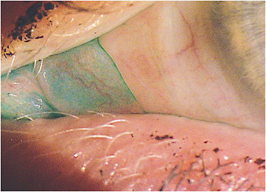
Lissamine Green in Dry Eye Practice
This image depicts diffuse lissamine green staining of the conjunctiva in a patient who has severe dry eye. It is worth noting that this area of damage did not stain with sodium fluorescein.
Vital dyes have been utilized to assess the integrity of the ocular surface for more than a century. Lissamine green is a vital dye, i.e., a staining agent used in the coloration of living cells or other constituents of tissue.1 M.S. Norn, a Danish researcher, evaluated the use of vital dyes on the ocular surface. In 1964, Norn described double-staining conjunctival tissue with rose bengal and alcian blue dyes.2 He later described the vital staining characteristics of lissamine green on the ocular surface.3 He noted that lissamine stains degenerate cells, dead cells, and mucus, and its vital‐staining properties are almost identical to those of rose bengal.3
Although rose bengal has been investigated as a diagnostic tool for evaluating dry eye, it differs from lissamine green in one important feature: comfort; subjects report that rose bengal is much more irritating when compared to lissamine green.4
Korb et al evaluated corneal and conjunctival staining characteristics as well as patient preference based on comfort between two preparations.5 The first was a combination of sodium fluorescein and rose bengal, and the second was sodium fluorescein and lissamine green. They found essentially identical staining characteristics between the two preparations, but all subjects preferred the lissamine green preparation due to significant discomfort associated with the rose bengal.5
Practitioner-based evaluation of ocular surface staining is somewhat subjective and can be dependent on illumination, the concentration of stain, and observer bias. Computer-based evaluation of imaging could be very beneficial in terms of time savings. Bunya et al obtained images of ocular lissamine green staining in 11 subjects. They compared the grading scores by two ophthalmologists using the van Bijsterveld scale and the National Eye Institutes (NEI) scale to computerized evaluation of the same images using the same scales. They found good correlation between the observers and computer evaluation in the van Bijsterveld scale but not in the NEI scale.6
Lissamine green is beneficial in the diagnosis and ongoing evaluation/management of patients suspected or diagnosed with dry eye. If it is not currently part of your dry eye protocols, consider adding it as part of your everyday regimen.
REFERENCES
- Begley C, Caffery B, Chalmers R, Situ P, Simpson T, Nelson JD. Review and analysis of grading scales for ocular surface staining. Ocul Surf. 2019 Apr;17:208-220.
- Norn MS. Specific double staining of the cornea and conjunctiva with rose bengal and alcian blue. Acta Ophthalmol (Copenh). 1964;42:84-96.
- Norn MS. Lissamine green. Vital staining of the cornea and conjunctiva. Acta Ophthalmol (Copenh). 1973;51(4):483-91.
- Manning FJ, Wehrly SR, Foulks GN. Patient tolerance and ocular surface staining characteristics of lissamine green versus rose bengal. Ophthalmology. 1995 Dec;102(12):1953-7.
- Korb DR, Herman JP, Finnemore VM, Exford JM, Blackie CA. An evaluation of the efficacy of fluorescein, rose bengal, lissamine green, and a new dye mixture for ocular surface staining. Eye Contact Lens. 2008 Jan;34:61-64.
- Bunya VY, Chen M, Zheng Y, et al. Development and Evaluation of Semiautomated Quantification of Lissamine Green Staining of the Bulbar Conjunctiva From Digital Images. JAMA Ophthalmol. 2017 Oct 1;135:1078–1085.


