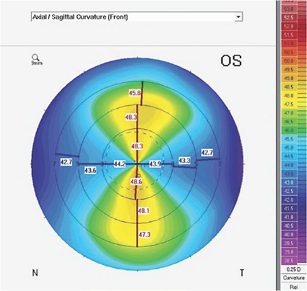
As practitioners expand their fitting skills and armamentarium, a deeper understanding of corneal topography mapping is beneficial. How can we better understand the differences in axial, tangential, and elevation maps, and how can this information enhance our fitting success?
Evaluating Different Topography Maps
Axial Maps Axial maps measure curvature of the anterior corneal surface using one radius of curvature for all points (Figure 1). Thus, axial maps are based on a sphere in which all points converge to a central point. Areas of greater curvature are shown in “warm” colors such as orange and red, whereas areas with flatter curves are illustrated in “cool” colors such as blue and violet. These maps average much of the data, resulting in simplification or smoothing. They are fairly simple to evaluate, and they indicate the orientation and degree of astigmatism present. This helps to separate corneal from internal astigmatism and to guide the lens recommendation (GP versus soft toric and appropriate initial diagnostic GP lens).

Tangential Maps Tangential or “instantaneous” maps use a number of radii to evaluate the curvature changes across the entire surface of the cornea (Figure 2). They utilize less “smoothing” and are “busier,” with more colors and detail of the entire corneal surface. Locating the cone apex in keratoconus may be more obvious than with an axial map, as more data are provided.

Elevation Maps Elevation maps show the high and low points of the corneal surface (Figure 3), which may be different from the areas of greatest curvature. Elevation maps are always in reference to the “best fit sphere” (BFS), which is an average of the corneal radius. Unlike axial and tangential maps, the warm, red-yellow colors indicate regions above the sphere—in other words, flatter—and the cooler, blue-violet colors indicate those areas below the BFS and therefore steeper. With study, these color differences become intuitive, much like interpreting the “upside-down-reverse” image that we see with binocular indirect ophthalmoscopy.

An elevation map is also useful when fitting scleral lenses in determining the location on the cornea where the lens is most likely to touch. When fitting a diagnostic lens, special attention may be directed to that area to ensure adequate clearance. Conversely, less elevated areas may have excessive clearance, necessitating a change in the base curve for a more uniform alignment.
With Scheimpflug imaging, both anterior and posterior elevations can be viewed. This is very useful in the early detection of keratoconus, which usually begins with an elevation of the posterior cornea. CLS


