A 45-year-old female presented for an annual exam. Her chief complaint was decreased near vision with her current glasses. She did not wear contact lenses and had no other symptoms of irritation or discomfort. Her history was only significant for a maternal family history of an inherited eye disease.
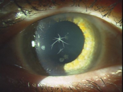
Her examination was uneventful with just a slight change in spectacle Rx and the corneal opacities observed in the photo (Figure 1). Her corneas did not stain with fluorescein or demonstrate any surface irregularity.

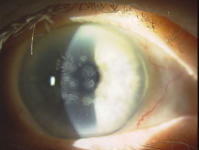
On careful observation of the opacities, they were found to be anterior stromal in location. As it turned out, the patient’s mother was also a patient of the clinic and had photos taken in the past as well (Figure 2).

After evaluation of the patient and her mother, who was previously diagnosed with granular corneal dystrophy, the patient was diagnosed with Avellino corneal dystrophy.
Avellino corneal dystrophy is an anterior stromal dystrophy that essentially comprises signs of both lattice type I dystrophy as well as granular type I dystrophy. Given that these dystrophies originate from mutations in the same gene, TGFBI on chromosome 5, it should not be surprising that there are cases that have signs of both the crumb-like flakes of granular type I, and the branching opacities of lattice type I. In this case, the mother manifests only signs of granular type I, while the daughter shows signs of both dystrophies, hence the categorization as Avellino corneal dystrophy.
Management of patients who have Avellino corneal dystrophy comes down to the symptoms that the patients manifest. These symptoms can range from mild irritation and discomfort to visual complications such as blur or glare. It is common for patients who have granular, lattice, and Avellino dystrophies to have very minimal symptoms with just mildly reduced visual acuity.
Patients who have irritation and discomfort can be managed with topical agents, such as artificial tears, gels, or, on occasion, a mild steroid. Visual symptoms can be managed with penetrating keratoplasty, deep anterior lamellar keratoplasty, or with photorefractive keratectomy.1,2 Laser-assisted in situ keratomileusis is not effective at managing visual symptoms and may in fact exacerbate the number and density of opacities.3 The opacities will recur in a cornea that has had surgical treatment, though the recurrence can take many years to develop. Soft contact lenses, particularly lower-Dk hydrogel lenses, can reduce the rate and risk of recurrence of opacities after surgical intervention.4
The patient in this instance had no real symptoms other than the mildest visual complaint, which did not warrant any intervention, only education. The remainder of her eye examination was unremarkable. The patient was reeducated as to the genetic aspects of the disease, prescribed new corrective lenses, and scheduled for her next annual exam.
References
- Kennedy SM, McNamara M, Hillery M, Hurley C, Collum LM, Giles S. Combined granular lattice dystrophy (Avellino corneal dystrophy). Br J Ophthalmol. 1996 May;80:489-490.
- Park SH, Mok J, Joo CK, Kim MS. Heterozygous Avellino corneal dystrophy 9 years after photorefractive keratectomy: natural or laser-induced accelerated course? Cornea. 2009 May;28:465-467.
- Jun RM, Tchah H, Kim TI, et al. Avellino corneal dystrophy after LASIK. Ophthalmology. 2004 Mar;111:463-468.
- Severin M, Konen W, Kirchhof B. Granular corneal dystrophy: treatment with soft contact lenses. Graefes Arch Clin Exp Ophthalmol. 1998 Apr;236:291-294.


