LET’S START with some advice: Practitioners should not forget about older technology and techniques in their toolbox. As new lens modifications are emerging, it is easy to forget about adjustments and systems that worked for them for years.
A 39-year-old male patient presented for a routine scleral contact lens follow-up complaining of increasing redness in his right eye that started one to two months ago. The patient had been lost to follow-up for months previously. His ocular history included keratoconus, previous hybrid contact lens wear for six years, and a recent switch to scleral contact lenses due to peripheral vascularization.
His last visit revealed that he was wearing a 17mm scleral lens and was noted to have 300 microns of central clearance, 100 microns of limbal clearance 360°, and mild inferior haptic impingement. Slit lamp exam documented a < 1mm hypertrophic conjunctival nodule next to the area of impingement. At that visit, the patient was asked to decrease wear time and was started on prednisolone acetate 1% ophthalmic suspension with the suspicion that this was a pyogenic granuloma due to mechanical rubbing from his lens.
The patient was then lost to follow-up for more than eight months and returned with complaints of redness OD. He reported that he tried prednisolone after his last visit and did not notice any improvement.
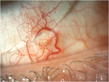
While being seen in the office, a 1.5mm hypertrophic conjunctival nodule with sectoral inflammation and injection on the bulbar conjunctiva was seen. Measurements and slit lamp photographs were taken. Measurements of the apex in relation to the edge of the lens, as well as the width, and an estimation of the height were noted. All other ocular findings were unchanged since his last visit. The decision to modify the lens incorporating toric haptics and a microvault were made (Figure 1).
Acquired epithelial inclusion cysts of the bulbar conjunctiva are formed when epithelial cells are displaced (Barnett and Johns, 2017). They are pushed into the substantia propria, where they proliferate forming a cyst cavity filled with clear liquid. Patients are usually asymptomatic.
In this case, the cyst was likely formed due to microtrauma from the inferior lens. There were a few ways this inclusion cyst could have been dealt with: 1) adjusting the overall diameter of the lens smaller or larger; 2) notch the lens to fit around the obstacle; 3) microvault; 4) impression molding technology or 5) switch to a rigid GP (Sherman et al, 2020).
In this case, a smaller lens was not chosen due to the patient’s advanced disease entity, and a larger lens was not possible due to the parameters available from the lens manufacturer. A notch was deferred due to the unpredictability of lens performance. The patient deferred impression molding technology due to cost. Ultimately, a microvault was chosen due the stability of our current lens.
The microvault is a local area of increased clearance that is created by a lathe (Barnett and Johns, 2017). It creates a designed flute in the edge of the scleral lens that vaults over the peripheral obstruction. This reduces the bearing over the area of concern and, therefore, decreases the irritation. The microvault is defined by its location, axis, decentration, width, and depth (Messer 2017). It works best in a very stable lens to not cause inflammation or desiccation of adjacent tissue.
In this case, fixing the acquired epithelial inclusion cyst took one visit and one new lens for the patient, which has now been stable for six years. CLS
References
- Barnett M, Johns LK. Contemporary Scleral Lenses: Theory and Application. Sharjah, United Arab Emirates, Bentham Science Publishers, 2017.
- Sherman SW, Cherny C, Suh LH. Epithelial Inclusion Cyst of the Bulbar Conjunctiva Secondary to Scleral Lens Impingement Managed With a MicroVault. Eye Contact Lens. 2020 Nov;46:e56-e58.
- Messer B. Utilizing MicroVaults to improve comfort and cosmesis in scleral lens wearers with pingueculae. Available at secure.toolkitfiles.co.uk/clients/24793/sitedata/PDF/Zenlens_Poster.pdf . Accessed Oct. 30, 2023.


