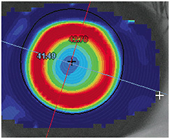
Keratoconus is a noninflammatory corneal disease that leads to stromal thinning, irregular astigmatism, high myopia, and impaired vision. This onset of corneal ectasia typically occurs in the second decade of life during puberty; however, pediatric cases have been reported as young as 4 years of age (Sabti et al, 2015).
Unfortunately, diagnosing children with keratoconus usually occurs after it has become advanced. This could be due to parents not knowing about the disease, ECPs having difficulty examining children, and/or ECPs not having the right corneal diagnostic equipment.
Corneal scarring is often seen in advanced stages, which is a risk for deprivation amblyopia in children. Although corneal transplantation can be performed to restore vision, children tend to have more postoperative complications such as graft dehiscence and failure, glaucoma, and early development of cataract (Huang et al, 2009).
Due to increasing awareness of the myopia epidemic, more children are being seen for eye exams, giving ECPs greater opportunity to examine for pediatric keratoconus.
HOW IT WORKS
Currently, a leading contact lens therapy for myopia management is orthokeratology.This technology utilizes a reverse geometry GP lens worn overnight to gently reshape the corneal surface, thereby correcting for daytime myopia. Understanding how ortho-k reshapes the corneal surface is foundational to detecting pediatric keratoconus.
In ortho-k, the cornea is primarily reshaped by movement of epithelial intracellular fluid from the central treatment zone into the adjacent reverse zone (Choo et al, 2008) (Figure 1). Early corneal changes during ortho-k change the epithelium. The epithelial layer of the cornea is approximately 50 microns thick centrally, and these cells are anteriorly joined by tight junctions, forming a continuous intercellular barrier (Sridhar, 2018). Posterior to the tight junctions, communication through gap junctions occurs in which intracellular fluid is exchanged between cells. It is this fluid, and the forces it transmits, that reshapes the cornea during orthokeratology (Choo et al, 2008).

Since there is a 50-micron limit to the thinning effects of ortho-k, pachymetry can be used to screen for pediatric keratoconus. Any corneal thinning beyond 50 microns should be suspicious for stromal thinning and indicates further testing for keratoconus.
If keratoconus is suspected during the initial myopia management evaluation, it may be best to discontinue ortho-k until keratoconus is ruled out. Beyond typical diagnostic criteria, a diagnosis of pediatric keratoconus is supported by results from genetic screening for corneal dystrophies, corneal slit lamp signs (e.g., Fleischer’s ring and Vogt’s striae), irregular astigmatism, and by posterior corneal evaluation via Scheimpflug tomography.
Summary
Myopia is increasingly common. Keratoconus is uncommon, but begins in childhood. Since ortho-k can exacerbate keratoconus, it is advisable to monitor corneal thickness during ortho-k myopia control exams. Early detection of keratoconus improves outcomes (Reinstein et al, 2015). An unexpected benefit of the myopia pandemic may be a reduced burden of advanced keratoconus in the future. CLS
References
- Sabti S, Tappeiner C, Frueh BE. Corneal Cross-Linking in a 4-Year-Old Child With Keratoconus and Down Syndrome. Cornea. 2015 Sep;34:1157-1160.
- Huang C, O’Hara M, Mannis MJ. Primary pediatric keratoplasty: indications and outcomes. Cornea. 2009 Oct;28:1003-1008.
- Choo JD, Caroline PJ, Harlin DD, Papas EB, Holden BA. Morphologic changes in cat epithelium following continuous wear of orthokeratology lenses: a pilot study. Cont Lens Anterior Eye. 2008 Feb;31:29-37.
- Sridhar MS. Anatomy of cornea and ocular surface. Indian J Ophthalmol. 2018 Feb;66:190–194.
- Reinstein DZ, Archer TJ, Urs R, Gobbe M, RoyChoudhury A, Silverman RH. Detection of Keratoconus in Clinically and Algorithmically Topographically Normal Fellow Eyes Using Epithelial Thickness Analysis. J Refract Surg. 2015 Nov; 31:736–744.


