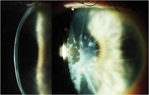

A 70-year-old male presented with reduced acuity and an inability to drive. He was 40 years post-radial keratotomy (RK) in both eyes and 10 years post cataract extraction with intraocular lens in both eyes. Entering vision was 20/300 OD and 20/150 OS. A refraction improved the vision to 20/100 in each eye.
Slit lamp examination revealed RK incisions in both eyes as well as a superficial, dense white plaque of deposited material in each eye that seemed to follow the corneal shape. Based on the appearance of the scar, the patient was diagnosed with a variant of a Salzmann’s type corneal degeneration, and he was fitted with scleral lenses to improve his acuity. While he tolerated the lenses well, his vision only improved to 20/70 and 20/60.
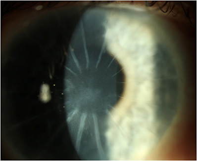
Due to the superficial nature of the opacity, the patient was offered a superficial keratectomy (SK) OD to attempt to improve his visual acuity. This was performed without complication; after re-epithelialization, his vision with his scleral lens improved to 20/20-2 (Figure 2).

Salzmann nodular degeneration typically presents as somewhat round, bluish-white elevated nodules on the corneal surface. These nodules may or may not break through Bowman’s layer. When they do not penetrate Bowman’s layer, a simple superficial keratectomy may be adequate to remove them when they impact visual acuity to a significant degree that the benefit outweighs the risk. When the nodules are deeper, a phototherapeutic keratectomy is preferred.
In this instance, due to the corneal shape change induced by the RK, the plaque of opacity formed a more diffuse, shallow pattern. The plaque was removed at the slit lamp under topical anesthetic with a spud tool and forceps. A bandage lens was placed over the cornea and the patient was treated with topical antibiotic drops for five days until re-epithelialization was complete. Once the bandage lens was removed, the patient resumed scleral lens wear and was pleased to report he was back to driving!


