CONTACT LENSES INHERENTLY disrupt the natural physiology of the anterior ocular surface, including decreasing the availability of atmospheric oxygen to the cornea during lens wear. This has the potential to create a hypoxic state that leads to complications including corneal vascularization and edema.
The oxygen permeability of the contact lens polymer, lens thickness, and oxygenated tear flow under the lens contribute the available oxygen to the cornea to maintain metabolic equilibrium. Changing lens design or surgery are sometimes necessary for specialty lens cases in which decreased endothelial cell function results in corneal edema secondary to scleral lens wear.
CASE #1
A 69-year-old female keratoconus patient, who had a left eye history of penetrating keratoplasty, Descemet stripping automated endothelial keratoplasty (DSAEK) surgery, and pseudophakia, was successfully wearing a 16mm scleral lens (tisilfocon A, Dk 180) with less than 100 microns of central corneal clearance, achieving 20/30 visual acuity. Her right eye had light-perception vision secondary to severe glaucoma.
After two episodes of corneal graft rejection, she started to develop corneal edema after two hours of scleral lens wear, resulting in poor vision. Her best-corrected visual acuity OS with refraction was 20/200 and, therefore, wearing a GP specialty lens was necessary for normal visual function.
Two attempts to fit her with corneal GP lenses to allow increased oxygenated tear flow to her cornea failed secondary to lens instability on her highly irregular corneal surface. Without other options, the patient had a repeat DSAEK surgery. Two months later, she restarted her previously designed scleral lens and, with a power adjustment, achieved 20/25 visual acuity.
CASE #2
A 64-year-old female, who was a lifelong glasses wearer, had cataract surgery in both eyes and subsequently complained of worse vision postoperatively with monocular diplopia. The patient was referred to our office and manifest refraction was OD –1.75 –1.25 x 138 20/70 and OS plano –0.75 x 092 20/60. Corneal topography revealed that she had keratoconus. Slit lamp exam was unremarkable. The patient was fit with 16.5mm scleral lenses (hexafocon A Dk 100) with less than 100 microns central corneal clearance, providing her with OD 20/25 and OS 30 visual acuity.
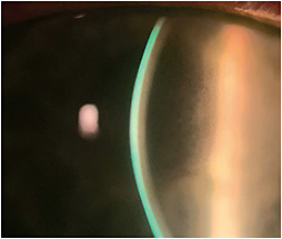
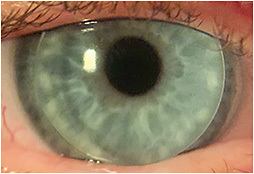
At follow-up, she reported that after several hours of lens wear, her vision appeared “steamy” with rainbows around lights, indicating lens-induced corneal edema (Figure 1). The patient was refit into 10mm corneal GP lenses (Figure 2) that allowed for tear exchange and prevented corneal edema with visual acuity of OD 20/20 and OS 20/25.


Significant corneal edema can result during scleral lens wear for corneas that have endothelial dysfunction, despite using hyper-Dk materials and minimized fluid reservoir thickness to maximize transmissibility. A study has shown that there is little benefit to adding a single fenestration to improve exchange (Fisher et al, 2022).
The case reports demonstrate that lamellar transplant surgery, such as DSAEK, to replace the endothelial layer or switching to a corneal GP that allows tear exchange are options for successful wear of a medically necessary GP lens. There is recent evidence that off-label use of rho-kinase inhibitors can help clear cornea edema (Davies, 2021) and potentially be used (off label) with scleral lens wear. CLS
References
- Fisher D, Collins MJ, Vincent SJ. Corneal oedema during open-eye fenestrated scleral lens wear. Ophthalmic Physiol Opt. 2022 Sep;42:1038-1043.
- Davies E. Case Series: Novel Utilization of Rho-Kinase Inhibitor for the Treatment of Corneal Edema. Cornea. 2021 Jan;40:116-120.


