IT IS COMMON for patients to require contact lenses following penetrating keratoplasty (PK) surgery, due to resultant irregular astigmatism, high refractive errors, and/or anisometropia (Wietharn and Driebe, 2004). It has been suggested that up to 47% of posttransplant eyes may require contact lens correction to achieve acceptable visual acuity (Brierly et al, 2000; Geerards et al, 2006).
However, fitting patients with contact lenses post-PK presents many challenges. The contour of the corneal graft is often highly irregular, making it difficult to achieve an acceptable lens fit. Additionally, transplant tissue often requires particular care in lens fitting to avoid certain complications that can lead to graft failure and/or rejection.
A 73-year-old white male presented with the complaint of vision issues with his corneal GP lens OD. He noted that his vision was good in the morning, but as the day went on, it started to get cloudy, with halos around lights. He had no symptoms OS.
His history included keratoconus, with PK OD in 1982 and OS in 2000, and successful GP lens wear ever since. On examination, he had a clear but decentered and tilted graft OD, with moderate peripheral vascularization. The graft OS was well centered and clear, with trace vascularization.
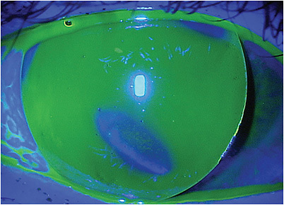
The fit of his GP lens OD wasn’t ideal (Figure 1), but multiple past efforts to refit him into different GP lens designs—both corneal and scleral—had been unsuccessful. He was resistant to fitting follow-ups and preferred a flat lens fit for easier lens removal, despite undesirable edge lift and the lens bearing inferiorly on a high point of the graft. The fit OS was reasonable. Though his right cornea appeared clear at the time of his exam, his symptoms suggested corneal edema, likely driven by an old graft stressed by hypoxia and decades of endothelial cell loss.

Corneal endothelial cell loss can be significant following PK and continues to occur throughout the life of the graft (Lass et al, 2011), so the risk of corneal edema is ever-present. It is critical to avoid inducing hypoxia with contact lenses, as this could serve as a compounding etiology for vascularization and edema, particularly in an at-risk cornea (Liesegang, 2002). This is perhaps another reason for this patient to stay in corneal GP lenses, as avoiding hypoxic conditions in scleral lenses can be difficult, even with significant modifications to the fit, such as minimizing vault and using hyper-Dk materials and smaller diameters (Compañ et al, 2014; Kumar et al, 2020; Michaud et al, 2012).
The patient’s habitual GP lens material had a Dk of 58 (ISO/Fatt). Fortunately, options in GP lens materials are now available that can provide more oxygen to compromised corneas than ever before. New lenses were ordered for both of the patient’s eyes with no change in parameters, but in a hyper-Dk GP material (tisilfocon A, Dk = 180). With this new material, the patient reported his symptoms resolved in full, and hopefully this will also prolong the life of his grafts. Utilizing these innovative materials can be very beneficial, even for corneal GP lens wearers. CLS
References
- Wietharn BE, Driebe WT Jr. Fitting contact lenses for visual rehabilitation after penetrating keratoplasty. Eye Contact Lens. 2004 Jan;30:31-33.
- Brierly SC, Izquierdo L Jr, Mannis MJ. Penetrating keratoplasty for keratoconus. Cornea. 2000 May;19:329-332.
- Geerards AJ, Vreugdenhil W, Khazen A. Incidence of rigid gas-permeable contact lens wear after keratoplasty for keratoconus. Eye Contact Lens. 2006 Jul;32:207-210.
- Lass JH, Beck RW, Benetz BA, et al; Cornea Donor Study Investigator Group. Baseline factors related to endothelial cell loss following penetrating keratoplasty. Arch Ophthalmol. 2011 Sep;129:1149-1154.
- Liesegang TJ. Physiologic changes of the cornea with contact lens wear. CLAO J. 2002 Jan;28:12-27.
- Compañ V, Oliveira C, Aguilella-Arzo M, Mollá S, Peixoto-de-Matos SC, González-Méijome JM. Oxygen diffusion and edema with modern scleral rigid gas permeable contact lenses. Invest Ophthalmol Vis Sci. 2014 Sep 4;55:6421-6429.
- Kumar M, Shetty R, Khamar P, Vincent SJ. Scleral Lens-Induced Corneal Edema after Penetrating Keratoplasty. Optom Vis Sci. 2020 Sep;97:697-702.
- Michaud L, van der Worp E, Brazeau D, Warde R, Giasson CJ. Predicting estimates of oxygen transmissibility for scleral lenses. Cont Lens Anterior Eye. 2012 Dec;35:266-271.


