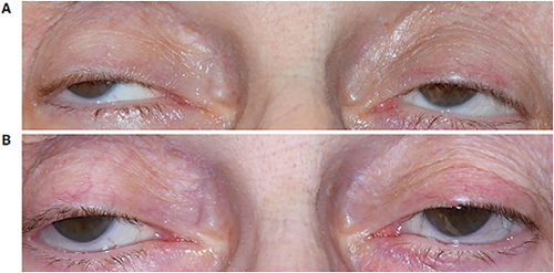
PATIENTS WITH PTOSIS are often told the options for improvement are blepharoplasty, spectacle crutches, eyelid tape, and/or therapeutic drops. The cause of ptosis can be myogenic, neurogenic, mechanical, or traumatic. While scleral lenses (SLs) typically are used for ocular surface disease or corneal disorders, an unconventional use is to improve ptosis.
CONTACT LENS OPTION
Historically, large-diameter SLs have been utilized for vaulting over advanced cones and/or proud corneal graphs. Another condition to add to this list is ptosis. A larger lens covers a greater surface area while increasing the central vault, resulting in an eyelid lift and ultimately improving lid cosmesis.
When fitting, manipulations to the diameter, base curvature, optic zone, and central clearance are pushed to the maximum parameters to gain the largest eyelid elevation.
CASE REPORTS
Severe chronic progressive ophthalmoplegia (CPEO) is a condition associated with ptosis. It is a progressive neuromyopathic disorder that can result in blepharoptosis and impaired ocular motility. The treatment can be extremely challenging because of the progressive nature of the disorder, making surgical intervention a poor option.
A 75-year-old-woman with a history of CPEO complained of constant brow elevation and chin lifting for visual improvement. The patient reported a history of failed ptosis crutches, and her oculoplastic surgeon refused to operate due to a probable poor long-term result.
On exam, the patient suffered from complete pupil coverage on both eyes, and the upper eyelids fell 2mm below the pupillary reflex (Figure 1A). The patient had a reduced interpalpebral fissure of 2mm OD and 1.5mm OS and was unable to move her extraocular muscles in any gaze of field. She was fit with SLs with an 18.5mm diameter, 8mm base curve, and 0.41 center thickness. At the final dispense visit, the edges of the SLs showed a flush landing with no signs of compression and around 350 microns of central clearance (Figure 1B).

While using the SLs, the patient’s interpalpebral fissure increased to 3mm in both eyes. She struggled with application and removal due to anxiety and poor dexterity; however, she overcame those obstacles.
In the literature, there have been limited cases of the use of SLs for ptosis correction. Katsoulos and colleagues (2018) tried high-vault SLs in the pediatric population, which showed increased upper eyelid support and widened palpebral aperture. Lindsay and Ezekiel (1997) reported using large-diameter SLs with ptosis props in a patient who had progressive bilateral ptosis due to ocular myopathy.
Cherny and colleagues (2021) reported a case of a 28-year-old patient with CPEO, however, due to the patient’s younger age the disease was less progressed. This patient got more than 3mm of improvement from SLs.
The patient in each of these cases varied in age, surgical history, and cause of the ptosis, which made it difficult to compare them directly, but practitioners and patients did report improvement in cosmesis, dry eye symptoms, visual acuity, and patient self-esteem.
Lens fitters looking for a nonsurgical, temporary alternative might try treating ptosis with large diameter SLs. CLS
References
- Katsoulos K, Rallatos GL, Mavrikakis I. Scleral contact lenses for the management of complicated ptosis. Orbit. 2018 Jun;37:201-207.
- Lindsay RG, Ezekiel DF. Ptosis prop gas permeable scleral lens fitting for a patient with ocular myopathy. Clin Exp Optom. 1997 Jul;80:123-126.
- Cherny C, Sherman SW, Dagi Glass LR. Severe chronic progressive external ophthalmoplegia-associated ptosis successfully treated with scleral lenses. J Neuroophthalmol. J Neuroophthalmol. 2021 Mar 1;41:137-138.


