RECURRENT CORNEAL EROSION (RCE) is characterized by repeated episodes of spontaneous breakdown of the corneal epithelium. Patients who have RCE experience foreign body sensation, pain, photophobia, and tearing (Brown and Bron, 1976). Also, clinical signs of RCE include loosely adherent and elevated epithelium, epithelial microcysts, corneal epithelial defects, stromal infiltrates and opacities, and epithelial basement membrane dystrophy (Brown and Bron, 1976).
The most frequent cause of RCE is mechanical trauma to the corneal surface with an object, such as a fingernail or piece of paper (Brown and Bron, 1976). Diabetes mellitus (Friend and Thoft, 1984) as well as a variety of corneal dystrophies have also been identified as risk factors for RCE (Hykin et al, 1994). A defect in epithelial anchoring is hypothesized as the cause of corneal epithelial erosions (Aitken et al, 1995).
RCE is usually treated in a stepwise fashion with the use of antibiotic lubrication drops and ointments. Medical soft bandage lenses can be applied to protect the affected cornea from the eyelid and to provide a healing scaffold (Wang and Jacobs, 2022). In acute RCE, bandage lenses can provide immediate relief (Wang and Jacobs, 2022). In some cases these conventional therapies are not effective, and debilitating symptoms persist for a long time. In recalcitrant cases, topical corticosteroids and oral doxycycline may also be helpful in healing defects associated with RCE (Dursun et al, 2001).
Surgical treatment such as anterior stromal micropuncture (Avni Zauberman et al, 2014) and excimer laser phototherapeutic keratectomy (Stasi and Chuck, 2009) may also be undertaken. Nevertheless, with proper implementation of medical bandage lenses and oral/topical therapies, surgery can be avoided (Wang and Jacobs, 2022).
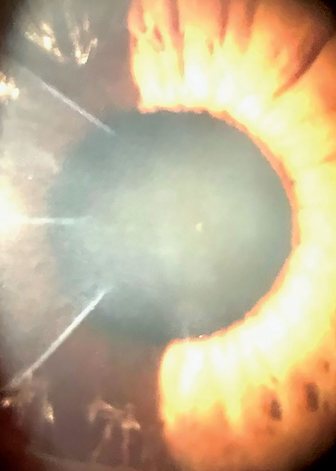
For example, a 72-year Caucasian female had a history of corneal scarring post radial keratectomy and basement membrane dystrophy (Figure 1). Upon wakening, the patient experienced abrupt episodes of severe pain in her left eye caused by RCE. Finally, the episodes were alleviated after she initiated artificial tears during waking hours and protection with a soft bandage daily disposable lens during sleep. Prophylactic antibiotic drops were instilled before each use of a bandage lens. After the eye healed, several attempts were made to discontinue soft lens therapy but were unsuccessful due to recurrence of pain.

To try to remove nighttime lens treatment, surgery was offered, but the patient declined. Her blurry vision due to irregular astigmatism was corrected with daytime scleral lens wear. The combination of topical therapy, nighttime soft bandage lens protection, and daytime scleral lens wear managed her RCE symptoms and rehabilitated her vision. The patient has been managed with this treatment regime for five years without complications.
REFERENCES
1. Brown N, Bron A. Recurrent erosion of the cornea. Br J Ophthalmol. 1976 Feb;60:84-96.
2. Friend J, Thoft RA. The diabetic cornea. Int Ophthalmol Clin. 1984;24:111-123.
3. Hykin PG, Foss AE, Pavesio C, Dart JK. The natural history and management of recurrent corneal erosion: a prospective randomised trial. Eye (Lond). 1994;8:35-40.
4. Aitken DA, Beirouty ZA, Lee WR. Ultrastructural study of the corneal epithelium in the recurrent erosion syndrome. Br J Ophthalmol. 1995 Mar;79:282-289.
5. Wang X, Jacobs DS. Contact Lenses for Ocular Surface Disease. Eye Contact Lens. 2022 Mar 1;48:115-118.
6. Dursun D, Kim MC, Solomon A, Pflugfelder SC. Treatment of recalcitrant recurrent corneal erosions with inhibitors of matrix metalloproteinase-9, doxycycline and corticosteroids. Am J Ophthalmol. 2001 Jul;132:8-13.
7. Avni Zauberman N, Artornsombudh P, Elbaz U, Goldich Y, Rootman DS, Chan CC. Anterior stromal puncture for the treatment of recurrent corneal erosion syndrome: patient clinical features and outcomes. Am J Ophthalmol. 2014 Feb;157:273-279.e1.
8. Stasi K, Chuck RS. Update on phototherapeutic keratectomy. Curr Opin Ophthalmol. 2009 Jul;20:272-275.


