


contact lens case reports
Software Aids in Lens Design for Post-LASIK Patient
BY PATRICK J. CAROLINE, FAAO, & MARK P. ANDRÉ, FAAO
March 2001
Patient N.O. is a 46-year-old female who underwent bilateral LASIK in 1998 for correction of 7.00D of myopia. Her post-operative refraction OD was +0.50 0.50 x 170 20/20 and OS +2.00 1.25 x5 20/20. The patient desired contact lens correction for her left eye for distance only. She was content with her reading glasses for near.
|
|
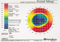
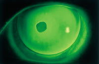
| Figure 1. Post-op map with a mid-peripheral radius of 43.50 diopters. | Figure 2. Diagnostic lens with a base curve of 43.50 diopters. |
The post-op cornea map OS revealed central K readings of 38.00 @ 6 / 39.78 @ 96. We selected a diagnostic lens with a base curve of 43.50, equal to the temporal radius of curvture along the horizontal meridian 4.0 from center (Figure 1). Slit lamp examination revealed mid-peripheral alignment, with a large fixed bubble over the central ablation zone (Figure 2).
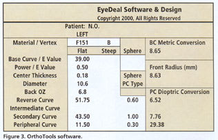
We designed a reverse geometry RGP lens using the OrthoTools software from EyeDeal Software in Fresno, CA. We entered into the program parameters such as base curve, power, diameter, optic zone, mid-peripheral fitting curve and peripheral curve. The software calculated the radius of the reverse curve, which in this case was 51.75 diopters (6.52mm) (Figure 3).
The final lens revealed excellent centration with a slight degree of central apical clearance and mid-peripheral alignment in the area of the fitting/secondary curve (Figure 4).
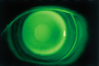
Figure 4. Reverse geometry lens with
a mid-peripheral radius of 43.50D.
Patrick Caroline is an associate professor of optometry at Pacific University and an assistant professor of ophthalmology at the Oregon Health Sciences University. Mark André is director of contact lens services at the Oregon Health Sciences University.