contact lens case reports
Fitting Reverse Geometry Lenses Post-ALK
BY PATRICK J. CAROLINE, FAAO, FCLSA, & MARK P. ANDRÉ,
FAAO, FCLSA
September 2001
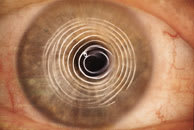
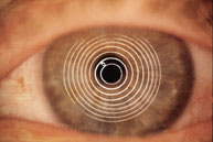
Patient D.P. underwent bilateral ALK in 1996 for correction of 9.00D. Photokeratoscopy of her right eye showed irregular astigmatism secondary to a microkeratome malfunction, with a best-corrected spectacle acuity of 20/60. The left eye showed regular corneal astigmatism with a refraction of 1.00 0.75 x 150 20/20 (Figure 1). So, let's design a reverse geometry lens OD.
|
|
|
| Figure 1a. Photokeratoscopy OD. | Figure 1b. Photokeratoscopy OS. |
|
|
|
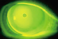
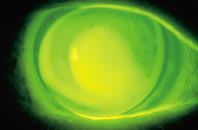
| Figure 2. A traditional RGP lens with excellent mid-peripheral alignment. | Figure 3. The reverse geometry lens on the patient's right eye. |
Step 1. Determine the central base curve radius. Clinical experience has taught us that an excellent starting base curve radius is 1.00D steeper than flat K. Because K readings and/or cor-neal mapping were not possible on our patient, we empirically selected a base curve radius of 8.60mm (39.25 diopters).
Step 2. Determine the mid-peripheral alignment curve radius. The alignment curve on a reverse geometry lens is the single lens parameter responsible for the fitting characteristics (centering and movement) of the lens. Determine its radius by corneal mapping and selecting a curve equal to the temporal mid-peripheral cornea 4.0mm from center. Alternately, you can determine the alignment curve radius with traditional diagnostic lenses. Insert an RGP lens with a base curve radius of 43.00 diopters and evaluate the mid-peripheral lens-to-corneal fitting relationship with fluorescein. We achieved mid-peripheral lens alignment with a radius of 44.00 diopters (7.67mm) (Figure 2).
|
|
|
|
Figure 4. A reverse curve is calculated to join the base curve with the alignment
curve. |
|
Step 3. Determine the reverse curve radius. The ultimate goal is a lens that significantly reduces the amount of apical clearance, yet provides mid-peripheral alignment for optimum lens centration, movement and comfort (Figure 3). The reverse curve joins the central base curve radius with the alignment curve radius (Figure 4). The laboratory or practitioner can make this calculation by incorporating sagittal height calculations into any number of software programs. In our case, the software indicated that a reverse curve radius of 6.58mm (51.25 diopters), would join two previously established radii.
Patrick Caroline is an associate professor of optometry at Pacific University and an assistant professor of ophthalmology at the Oregon Health Sciences University.
Mark André is director of contact lens services at the Oregon Health Sciences University.


