orthokeratology today
Ortho-k Success:
It's All in the Follow Up
BY JOHN MARK JACKSON, OD, MS, FAAO
Much of the clinical skill required for orthokeratology occurs during the lens fitting process -- but the lens fit is only the beginning of effective ortho-k treatment. Proper follow-up care after the fit is necessary to achieve a successful outcome.
Starting with Day One
Schedule a patient's first visit for the morning after he begins overnight lens wear. Many clinicians ask the patient to wear the lenses to the office, assuming good acuity with the lenses. You may lose valuable fitting information if the patient removes the lenses before he arrives because the impact that the lenses have on topography or acuity is most observable upon lens removal.
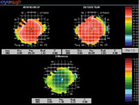
Include in your exam unaided acuities, refraction, topography and slit lamp findings. First-day treatment responses vary among patients. Continue lens wear if the topography shows a well-cen tered treatment zone with reduced refractive error (Figure 1) and no central island (Figure 2). Discontinue lens wear and let the cornea return to baseline before refitting if any undesirable topography patterns occur. Likewise, if the cornea shows edema or ex cessive staining, then discontinue lens wear until you address these issues.

|

|
Figure 1. Ideal first-day topography. |
Figure 2. Central island pattern. |
Monitoring Initial Changes
Check unaided acuities, refraction, topography and ocular health at one week. By this time the patient should be doing well -- if the lenses are correct. Don't make any changes if you've noticed steady improvement, even if it's not optimal. Change the lens only if minimal progress has occurred.
Looking for Stabilization
At two weeks, check unaided acuities, refraction, topography and slit lamp findings. At this time the cornea has typically changed as much as it can with a particular pair of lenses. The patient should function well without correction; if not, then change the lenses. Flatten the base curve to achieve more correction if his undercorrection is less than 1.00D. The sag depth is too deep if his undercorrection is more than 1.00D, so flatten the reverse curve, alignment curve or both.
Let the Patient Guide You
After two weeks, the follow-up schedule depends on how well the patient has responded. If you did not make any lens adjustments, then schedule visits at one, three, six and 12 months to monitor the patient's corneal health and visual function. If you made adjustments to the lenses, then schedule more frequent visits until you achieve a maximum change.
Dr. Jackson is an assistant professor at Southern College of Optometry where he works in the Advanced Contact Lens Service, teaches courses in contact lenses and performs clinical research.


